Expansion Microscopy for Cell Biology
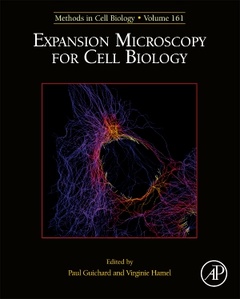
1. Protein-retention Expansion Microscopy: Improved Sub-cellular Imaging Resolution through Physical Specimen Expansion Paul Tilberg 2. Ultrastructure Expansion Microscopy (U-ExM) Davide Gambarotto, Virginie Hamel and Paul Guichard 3. Expansion STED microscopy (ExSTED) Helge Ewers 4. Simple multi-color super-resolution by X10 microscopy Sven Truckenbrodt 5. Expansion microscopy imaging of various neuronal structures Jae-Byum Chang 6. Mapping the neuronal cytoskeleton using expansion microscopy Lukas Kapitein 7. Mechanical expansion microscopy Bo Wang 8. Enhanced expansion microscopy to measure nanoscale structural and biochemical remodelling in single cells Izzy Jayasinghe 9. Application of Expansion Microscopy on Developing Arabidopsis Seeds Michael D. Nodine 10. A protocol to expand plant nuclei Veit Schubert 11. Expansion microscopy of the mitotic spindle Ivana Ponjavic 12. Expansion microscopy at the nanoscale. The nuclear pore complex as fiducial landmark Paolo Bianchini 13. Post-labeling expansion microscopy, a promise to go beyond super-resolution limitations Paul Guichard and Virginie Hamel 14. Ex-dSTORM and automated quantitative image analysis of expanded filamentous structures Markus Sauer 15. Expansion Microscopy on Drosophila Spermatocyte Centrioles Alan Wainman
Researchers in the area of Cell biology, Plant biology, Neurobiology as well as other scientific areas.
- Provides the authority and expertise of leading contributors from an international board of authors
- Represents the latest release in the Methods in Cell Biology series
- Includes the latest information on Expansion Microscopy for Cell Biology
Date de parution : 01-2021
Ouvrage de 356 p.
19x23.3 cm
Thème d’Expansion Microscopy for Cell Biology :
Mots-clés :
Airyscan; Anti-photobleaching; Arabidopsis thaliana; Asterless; Bacteria; Biochemical features; Bridging fiber; Cardiomyocytes; Cell wall; CENH3; Centriole; Centrioles; Connectivity; Cytoskeleton; DNA-antibody conjugation; Drosophila; dSTORM; Embryogenesis; Enhanced expansion microscopy; Epitope accessibility; Expansion; Expansion microscopy; Expansion microscopy (ExM); ExSTED; Fluorescence microscopy; Fluorescent immunostaining; Fluorescent proteins; Gel embedding; Hippocampal neurons; Hutchinson-Gilford progeria syndrome; Immunofluorescence; Immuno-staining; Interpenetrating polymer networks; Isolated nuclei; Isotropicity; Iterative expansion microscopy (iExM); Kinetochore fiber; LineProfiler; Linkage error; Macromolecular assemblies; Mechanically locked expansion microscopy; Mechanically resolved expansion microscopy; Microscopy; Microtubule; Microtubules; Mitotic spindle; Nanodomains; Nanoscale; Nervous system; Neurobiology; Nuclear lamina; Nuclear pore complex; Nucleoplasmic reticulum; Nup153; Planarian flatworm; Plant chromatin; Post-expansion labeling; PRC1; Rabl configuration; Re-embedding; Remodeling; Single cells; Spermatocyte; STED; Stimulated emission depletion microscopy; Structural resolution; Structured illumination microscopy; Super-resolution; Super-resolution fluorescence imaging; Super-resolution microscopy; Synapses; Synaptic vesicles; Tissue clearing; Tubulin; U-ExM; Ultrastructure expansion microscopy; X10; X10 expansion microscopy