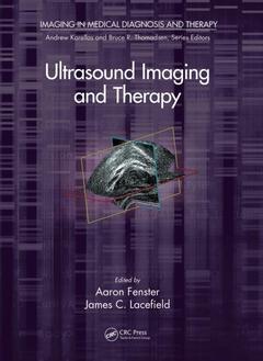
Ultrasound Imaging and Therapy Imaging in Medical Diagnosis and Therapy Series
Coordonnateurs : Fenster Aaron, Lacefield James C.
Up-to-Date Details on Using Ultrasound Imaging to Help Diagnose Various Diseases
Due to improvements in image quality and the reduced cost of advanced features, ultrasound imaging is playing a greater role in the diagnosis and image-guided intervention of a wide range of diseases. Ultrasound Imaging and Therapy highlights the latest advances in using ultrasound imaging in image-guided interventions and ultrasound-based therapy. The book presents current and emerging techniques, identifies trends in the use of ultrasound imaging, and addresses technical and computational problems that need to be solved.
The book is organized into three sections. The first section covers advances in technology, including transducers (2-D, 3-D, and 4-D), beamformers, 3-D imaging systems, and blood velocity estimation systems. The second section focuses on diagnostic applications, such as elastography, quantitative techniques for therapy monitoring and diagnostic imaging, and ultrasound tomography. The final section explains the use of ultrasound in image-guided interventions for image-guided biopsy and brain imaging.
ULTRASOUND INSTRUMENTATION: Array Transducers and Beamformers. Three-Dimensional Ultrasound Imaging, Ultrasound Velocity Imaging. DIAGNOSTIC ULTRASOUND IMAGING: Ultrasound Elastography. Quantitative Ultrasound Techniques for Diagnostic Imaging and Monitoring of Therapy. Ultrasound Tomography: A Decade-Long Journey from the Laboratory to the Clinic. Task-Based Design and Evaluation of Ultrasonic Imaging Systems. Acoustic Radiation Force–Based Elasticity Imaging. THERAPEUTIC AND INTERVENTIONAL ULTRASOUND IMAGING: Three-Dimensional Ultrasound-Guided Prostate Biopsy. Ultrasound Applications in the Brain. Index.
Aaron Fenster, PhD, is a founding director of the Imaging Research Laboratories at the Robarts Research Institute. He is also a professor in the Department of Medical Biophysics and Department of Medical Imaging and the founder and associate director of the Graduate Program in Biomedical Engineering at the University of Western Ontario. His research focuses on the development of 3-D ultrasound imaging with diagnostic, surgical, and therapeutic cancer applications.
James C. Lacefield, PhD, is an associate professor in the Department of Electrical and Computer Engineering and Department of Medical Biophysics, a faculty member of the Graduate Program in Biomedical Engineering, and a mentor in the CIHR Strategic Training Program in Cancer Research and Technology Transfer at the University of Western Ontario. He is also an associate scientist of the Imaging Research Laboratories at Robarts Research Institute. His research activities address physical acoustics and signal processing aspects of biomedical ultrasound imaging, with an emphasis on applications of ultrasound to cancer and cardiovascular research.
Date de parution : 05-2015
17.8x25.4 cm
Date de parution : 04-2018
17.8x25.4 cm
Thèmes d’Ultrasound Imaging and Therapy :
Mots-clés :
Ch Ap; Elasticity Imaging; aaron; QUS Parameter; fenster; ARFI Imaging; elasticity; TRUS Image; shear; TRUS Guide Prostate Biopsy; waves; Prostate Biopsy; acoustic; Wiener Filter; impedance; Shear Wave; prostate; Sound Speed; biopsy; TRUS Probe; ultrasonic; RF Data; Ideal Observer; SSI; Large Aperture Arrays; Strain Images; ARFI; Manual Segmentations; RF Echo; MF; High Intensity FUS; Biopsy Core; Inverse Elasticity Problem; Grating Lobes; Lumen Boundaries