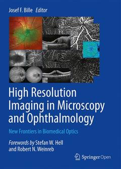
High Resolution Imaging in Microscopy and Ophthalmology, 1st ed. 2019 New Frontiers in Biomedical Optics

This open access book provides a comprehensive overview of the application of the newest laser and microscope/ophthalmoscope technology in the field of high resolution imaging in microscopy and ophthalmology. Starting by describing High-Resolution 3D Light Microscopy with STED and RESOLFT, the book goes on to cover retinal and anterior segment imaging and image-guided treatment and also discusses the development of adaptive optics in vision science and ophthalmology. Using an interdisciplinary approach, the reader will learn about the latest developments and most up to date technology in the field and how these translate to a medical setting.
High Resolution Imaging in Microscopy and Ophthalmology ? New Frontiers in Biomedical Optics has been written by leading experts in the field and offers insights on engineering, biology, and medicine, thus being a valuable addition for scientists, engineers, and clinicians with technical and medical interest who would like to understand the equipment, the applications and the medical/biological background.
Lastly, this book is dedicated to the memory of Dr. Gerhard Zinser, co-founder of Heidelberg Engineering GmbH, a scientist, a husband, a brother, a colleague, and a friend.
PART ONE - Breaking the Diffraction Barrier in Fluorescence Microscopy.- High-Resolution 3D Light Microscopy with STED and RESOLFT.- PART TWO - Retinal Imaging and Image Guided Retina Treatment.- Scanning Laser Ophthalmoscopy (SLO).- Optical Coherence Tomography (OCT) -Principle and Technical Realization.- Ophthalmic Diagnostic Imaging – Retina.- Ophthalmic Diagnostic Imaging – Glaucoma.- OCT Angiography (OCTA) in Retinal Diagnostics.- OCT-based Velocimetry for Blood Flow Quantification.- In Vivo FF-SS-OCT Optical Imaging of Physiological Responses to Photostimulation of Human Photoreceptor Cells.- Two-Photon Laser Scanning Ophthalmoscope.- Fluorescence Lifetime Imaging Ophthalmoscopy (FLIO).- Selective Retina Therapy.- PART THREE - Anterior Segment Imaging and Image Guided Treatment.- In Vivo Confocal Scanning Laser Microscopy.- Anterior Segment OCT.- Femtosecond-Laser-Assisted Cataract Surgery (FLACS).- Refractive Index Shaping – In-Vivo Optimization of an Implanted Intraocular Lens (IOL).- PART FOUR- Adaptive Optics in Vision Science and Ophthalmology.- The Development of Adaptive Optics and its Application in Ophthalmology.- Adaptive Optics for Photoreceptor-Targeted Psychophysics.- Compact Adaptive Optics Scanning Laser Ophthalmoscope with Phase Plates.
Professor Josef Bille of the University of Heidelberg in Germany is a pioneer in the area of laser eye correction. He developed a method for mapping irregularities in the cornea with unprecedented precision and fine-tuning the lasers to repair them. This ground-breaking invention, and its continuous improvement, has corrected near-sightedness, far-sightedness, and astigmatism in millions of patients worldwide.
Date de parution : 09-2019
Ouvrage de 407 p.
17.8x25.4 cm


