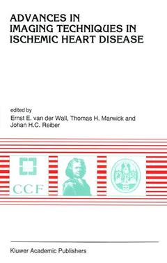
Advances in Imaging Techniques in Ischemic Heart Disease, Softcover reprint of the original 1st ed. 1995 Developments in Cardiovascular Medicine Series, Vol. 171
Langue : Anglais
Coordonnateurs : van der Wall Ernst E., Marwick Thomas H., Reiber Johan H. C.

In recent years there have been tremendous advances in cardiac imaging techniques covering the complete spectrum from echocardiography, nuclear cardiology, magnetic resonance imaging to contrast angiography. With respect to these noninvasive and invasive cardiac imaging modalities, marked technological developments have allowed the cardiologist to visualize the myocardium in a far more refined manner than conventional imaging was capable of. Echocardiography has extended its domain with intravascular ultrasound, cardiovascular nuclear imaging has added positron emission tomography to its line of research, magnetic resonance imaging has been broadened with magnetic resonance angiography and spectroscopy, and finally contrast angiograp hy has widened its scope with excellent quantitation programs. For all these imaging modalities it is true that the application of dedicated quantitative analytic software packages enables the evaluation of the imaging studies in a more accurate, reliable, and reproducible manner. It goes without saying that these extensions and achievements have resulted in improved diagnostics and subsequently in improved patient care. Particularly in patients with ischemic heart disease, major progress has been made to detect coronary artery disease in an early phase of the disease process, to follow the atherosclerotic changes in the coronary arteries, to establish the functional and metabolic consequences of the luminal obstructions, and to accurately assess the results of interventional therapy.
List of contributors. Preface. Current status of myocardial perfusion scintigraphy; E.E. van der Wall. Use of positron emission tomography for the diagnosis and evaluation of ischemic heart disease; Th.H. Marwick. Magnetic resonance coronary angiography; P.M.T. Pattynama, A. de Roos. Noninvasive imaging of coronary artery anomalies. Present angiographic criteria and role of additional techniques, especially fast gradient echo magnetic resonance imaging; H.W. Vliegen, et al. Left ventricular function by stress MR imaging. Animal studies using dobutamine; J. Baan, P.M.T. Pattynama. Spectroscopy of the human heart: techniques, limitations and opportunities; P.R. Luyten. Current status of stress echocardiography for the diagnosis of myocardial ischemia and viability; Th.H. Marwick. Contrast ultrasound for assessment of myocardial perfusion: promise and pitfalls; J.D. Thomas. Intravascular ultrasound. Possibilities of image enhancement by signal processing; N. Bom, et al. Clinical aspects of intravascular ultrasound; E.M. Tuzcu. Evolution of quantitative coronary arteriography; J.H.C. Reiber, et al. Index.
Date de parution : 10-2012
Ouvrage de 159 p.
16x24 cm
Disponible chez l'éditeur (délai d'approvisionnement : 15 jours).
Prix indicatif 105,49 €
Ajouter au panierThème d’Advances in Imaging Techniques in Ischemic Heart Disease :
© 2024 LAVOISIER S.A.S.


