Atlas of the Human Body Central Nervous System and Vascularization
Auteurs : Vidic Branislav, Milisavljevic Milan
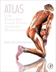
Atlas of Human Body: Central Nervous System and Vascularization is a multidisciplinary approach to the technical coverage of anatomical structures and relationships. It contains surface and 3D dissection images, native and colored cross sectional views made in different planes, MRI comparisons, demonstrations of cranial nerve origins, distribution of blood vessels by dissection, and systematic presentation of arterial distribution from the precapillary level, using the methyl metacrylate injection and subsequent tissue digestion method.
Included throughout are late prenatal (fetal) and early postnatal images to contribute to a better understanding of structure/relationship specificity of differentiation at various developmental intervals (conduits, organs, somatic, or branchial derivatives). Each chapter features clinical correlations providing a unique perspective of side-by side comparisons of dissection images, magnetic resonance imaging and computed tomography. Created after many years of professional and scientific cooperation between the authors and their parent institutions, this important resource will serve researchers, students, and doctors in their professional work.
1. Upper Limb and Vascularization2. Lower Limb and Vascularization3. Thorax and Vascularization4. Abdomen and Vascularization5. Pelvis and Perineum with 5–6-Month-Old Fetal Specimens6. Head and Neck Regions and Vascularization7. Cranial Central Nervous System and Spinal Cord8. Vascularization of Head and Neck and the Cranial Central Nervous System
Medical researchers of human physiology, student body and faculty of Medical and Paramedical Institutions, postdoctoral Fellows in residency, practicing Physicians and Surgeons
Dr. Vidic is an active member of many committees including:
• First Year Curriculum
• Scientific Merit
• Integration of Preclinical Instructions
• Faculty Promotion
• Equipment
• Correlation Conferences
• Educational Policy
• Audiovisual Facilities
• Coordinator of Gross Anatomy Curriculum
• In charge of Electron Microscopical Facilities
• Graduate French Language Requirement
• Interviewer for Incoming Medical Students
• Committee on Admissions, School of Medicine
• Curriculum on Aging
• Dr. Raj Bhussry Memorial Fellowship
• Search Committee, Department of Anatomy
• Coordinator of Graduate Program (Master in Anatomy and Certificate in Occlusion).
• Committee on Education, School of Dentistry
• Chairman Anatomical Board of the District of Columbia
• Auditing Committee AAA
• Committee Graduate Thesis
• Coordinator Direction of Departmental Development Committee
• Space Committee
• Financial Committee
Dr. Vidic has an impressive list of Professional Memberships including:
• American Association of Anatomists
• American Society for Cell Biology
• Washington Electron Microscopical Society
• Society of the Sigma Xi
• Southern Society of Anatomists
• New York Academy of Science
• Cajal Club
• Yugoslave Society of Anatomists
• German Society of Anatomists
• Electron Microscopic Society of America
• American Thoracic Society
• French Association of Anatomists
• American Association for the Advancement of Science
• The Society for Quantitative Morpholo
- Contains over 700 color photos of ideal anatomical preparations and sections of each part of the body that have been prepared, recorded, and processed by the authors
- Covers existing gaps including developmental and prenatal periods, detailed vascular anatomy, and neuro anatomy
- Features a comprehensive alphabetical index of structures for ease of use
- Features a companion website which contains access to all images within the book
Date de parution : 05-2017
Ouvrage de 278 p.
21.4x27.6 cm