Scanning Electron Microscopy for the Life Sciences Advances in Microscopy and Microanalysis Series
Langue : Anglais
Coordonnateur : Schatten Heide
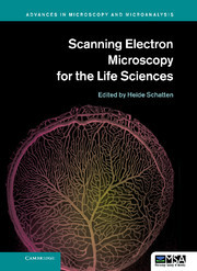
A guide to modern scanning electron microscopy instrumentation, methodology and techniques, highlighting novel applications to cell and molecular biology.
Recent developments in scanning electron microscopy (SEM) have resulted in a wealth of new applications for cell and molecular biology, as well as related biological disciplines. It is now possible to analyze macromolecular complexes within their three-dimensional cellular microenvironment in near native states at high resolution and to identify specific molecules and their structural and molecular interactions. New approaches include cryo-SEM applications and environmental SEM (ESEM), staining techniques and processing applications combining embedding and resin-extraction for imaging with high resolution SEM, and advances in immuno-labeling. New developments include helium ion microscopy, automated block-face imaging combined with serial sectioning inside an SEM chamber, and Focused Ion Beam Milling (FIB) combined with block-face SEM. With chapters written by experts, this guide gives an overview of SEM and sample processing for SEM and highlights several advances in cell and molecular biology that greatly benefited from using conventional, cryo, immuno and high-resolution SEM.
1. The role of scanning electron microscopy in cell and molecular biology: SEM basics, past accomplishments and new frontiers Heide Schatten; 2. Corrosion casting technique Jerzy Walocha, Jan A. Litwin and Adam J. Miodoński; 3. Revealing the internal structure of cells in three dimensions with scanning electron microscopy Sol Sepsenwol; 4. Mitochondria form continuous intracellular network-structures visualized with high-resolution field-emission scanning electron microscopy T. Naguro, H. Nakane and S. Inaga; 5. Chapter on 3-D reconstruction of cell organelles using STEM tomography Paul Walther; 6. High resolution labeling for correlative microscopy Ralph Albrecht, Daryl A. Meyer and O. E. Olorundare; 7. The use of SEM to explore virus structure and trafficking Jens M. Holl and Elizabeth R. Wright; 8. High resolution scanning electron microscopy of the nuclear surface in Herpes Simplex Virus 1 infected cells Peter Wild, Andres Kaech and Miriam S. Lucas; 9. Scanning electron microscopy of chromosomes: structural and analytical investigations Elizabeth Schroeder-Reiter and Gerhard Wanner; 10. A method to visualize the microarchitecture of glycoprotein matrices with scanning electron microscopy Giuseppe Familiari, Rosemarie Heyn, Luciano Petruzziello and Michela Relucenti; 11. Scanning electron microscopy of cerebellar intrinsic circuits Orlando J. Castejón; 12. Application of in vivo cryotechnique to living animal organs examined by scanning electron microscopy Shinichi Ohno, Nobuo Terada, Nobuhiko Ohno and Yasuhisa Fujii; 13. SEM in dental research Vladimir Dusevich, Jennifer R. Melander and J. David Eick; 14. SEM, teeth and palaeoanthropology: the secret of ancient human diets Alejandro Romero and Joaquín De Juan.
Heide Schatten is a Professor at the University of Missouri, Columbia. Her publications include advanced imaging methods, cellular and molecular biology, cancer biology, reproductive biology, microbiology and space biology. The latter included collaborations with NASA scientists and experiments aboard the Space Shuttle Endeavour to examine the effects of spaceflight on cytoskeletal organization during development. She has received numerous awards including grant awards from NASA, NIH and NSF. She has published over 185 papers, seven book chapters and edited several special topic journal issues and eight books with several more in progress.
Date de parution : 12-2012
Ouvrage de 298 p.
17x24.4 cm
Disponible chez l'éditeur (délai d'approvisionnement : 14 jours).
Prix indicatif 145,68 €
Ajouter au panierThèmes de Scanning Electron Microscopy for the Life Sciences :
© 2024 LAVOISIER S.A.S.