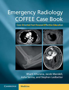
Emergency Radiology COFFEE Case Book Case-Oriented Fast Focused Effective Education
Langue : Anglais
Coordonnateurs : Khurana Bharti, Mandell Jacob, Sarma Asha, Ledbetter Stephen
This book of 85 index cases is organized by clinical presentations that simulate real-life radiology practice in the emergency department.
Emergency radiology requires consistent, timely, and accurate imaging interpretation with the rapid application of clinical knowledge across many areas of radiology practice that have traditionally been fragmented by organ system or modality divisions. This text unifies this body of knowledge into an educational resource capturing the core competencies required of an emergency radiologist. This book of 85 index cases is organized by clinical presentations that simulate real-life radiology practice in the emergency department. Companion cases spanning the differential diagnoses and spectrum of disease provide hundreds more examples for a fast, focused and effective education called COFFEE (Case-Oriented Fast Focused Effective Education). This text can serve as a 'go to' resource for radiologists, as well as any other physicians working in the emergency department. It will be an excellent companion for radiologists preparing for initial board certification or re-certification by the American Board of Radiology.
Part I. Non-Traumatic Conditions: Section 1. Abdomen: Section 1.1. Upper Quadrant and Epigastric Pain: Case 1. Acute cholecystitis; Case 2. Ruptured hepatic adenoma; Case 3. Acute pancreatitis; Case 4. Bile leak after cholecystectomy; Case 5. Splenic infarction; Case 6. Perforated duodenal ulcer; Section 1.2. Flank Pain: Case 7. Obstructive uropathy; Case 8. Retroperitoneal hematoma; Case 9. Bleeding renal angiomyolipoma; Section 1.3. Lower Quadrant Pain: Case 10. Appendicitis; Case 11. Crohn's disease; Case 12. Pseudomembranous colitis; Case 13. Sigmoid colon diverticulitis; Case 14. Meckel's diverticulitis; Case 15. Epiploic appendagitis; Section 1.4. Generalized Abdominal Pain: Case 16. Small bowel obstruction; Case 17. Internal hernia; Case 18. Foramen of Winslow hernia; Case 19. Obturator hernia; Case 20. Cecal volvulus; Case 21. Bowel perforation; Case 22. Ischemic colitis; Section 1.5. Pelvic/Scrotal Pain: Case 23. Tubo-ovarian abscess; Case 24. Ectopic pregnancy; Case 25. Gonadal vein thrombosis; Case 26. Ovarian torsion; Case 27. Fournier's gangrene; Case 28. Testicular malignancy; Case 29. Testicular torsion; Case 30. Renal vein thrombosis; Section 2. Thorax: Section 2.1. Chest Pain: Case 31. Pulmonary embolism; Case 32. Esophageal rupture; Case 33. Aortic dissection; Section 2.2. Cough/Shortness of Breath: Case 34. Pneumonia; Case 35. Empyema; Case 36. Lung abscess; Case 37. Lung cancer; Case 38. Pneumocystis pneumonia; Section 3. Otolaryngology: Case 39. Foreign body; Case 40. Peritonsillar abscess; Case 41. Sialolithiasis; Case 42. Sinusitis; Case 43. Longus coli calcific tendinopathy; Section 4. Neurology: Case 44. Stroke; Case 45. Glioblastoma multiforme; Case 46. Creutzfeldt-Jakob disease; Case 47. Intracranial hypotension; Case 48. Herpes Simplex virus encephalopathy; Case 49. Dural sinus thrombosis; Case 50. Subarachnoid hemorrhage; Case 51. Intraparenchymal hemorrhage; Case 52. Reversible vasoconstriction syndrome; Section 5. Musculoskeletal: Case 53. Spondylodiscitis; Case 54. Necrotizing fasciitis; Part II. Traumatic Conditions: Section 6. Neurology: Case 55. Diffuse axonal injury; Case 56. Epidural hematoma; Case 57. Vertebral artery dissection; Section 7. Thorax: Case 58. Blunt chest trauma; Section 8. Abdomen: Case 59. Diaphragmatic rupture; Case 60. Bladder rupture; Case 61. Liver trauma; Case 62. Splenic trauma; Case 63. Renal injury; Case 64. Ureteral trauma; Case 65. Uterine trauma; Section 9. Musculoskeletal: Section 9.1. Axial Skeleton Trauma: Case 66. Hyperflexion teardrop; Case 67. Hyperflexion distraction; Case 68. Pelvic trauma; Section 9.2. Upper Extremity Trauma: Case 69. Bennett fracture; Case 70. Monteggia fracture-dislocation; Case 71. Posterior elbow dislocation; Case 72. Proximal humeral fracture; Case 73. Radial head fracture; Case 74. Sternoclavicular dislocation; Case 75. Volar plate avulsion; Case 76. Acromioclavicular separation; Case 77. Anterior shoulder dislocation; Case 78. Distal radius fracture; Section 9.3. Lower Extremity Trauma: Case 79. Atypical femoral fracture; Case 80. Segond fracture; Case 81. Tibial plateau fracture; Case 82. Ankle supination external rotation injury; Case 83. Lisfranc ligament injury; Case 84. Fifth metatarsal fracture; Case 85. Sesamoid fracture; Index.
Bharti Khurana MD is Director of the Emergency Radiology Fellowship at Brigham and Women's Hospital and Assistant Professor of Radiology at Harvard Medical School, Massachusetts.
Jacob Mandell MD is a Fellow in Musculoskeletal Imaging and Intervention, class of 2014, and graduated resident in Diagnostic Radiology at Brigham and Women's Hospital and Harvard Medical School, Massachusetts.
Asha Sarma MD is a Diagnostic Radiology Chief Resident at Brigham and Women's Hospital and Harvard Medical School, Massachusetts.
Stephen Ledbetter MD MPH is Chief of Radiology at Brigham and Women's Faulkner Hospital, former Section Chief of Emergency Radiology at Brigham and Women's Hospital and Assistant Professor of Radiology at Harvard Medical School, Massachusetts.
Date de parution : 04-2016
Ouvrage de 672 p.
19x24.6 cm
Thème d’Emergency Radiology COFFEE Case Book :
© 2024 LAVOISIER S.A.S.