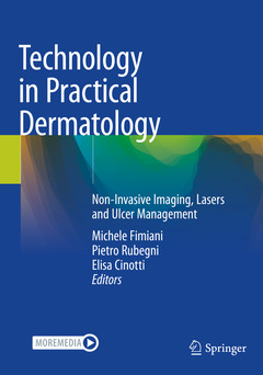
Technology in Practical Dermatology, 1st ed. 2020 Non-Invasive Imaging, Lasers and Ulcer Management

This book provides a complete overview on the latest available technologies in dermatology, while discussing future trends of this ever-growing field. This handy guide provides clinicians and researchers with a clear understanding of the advantages and challenges of laser and imaging technologies in skin medicine today. It also includes a section on imaging techniques for the evaluation of skin tumors, with chapters devoted to dermoscopy, in vivo and ex vivo reflectance confocal microscopy, high frequency ultrasound, optical coherence tomography, and a closing part on latest approaches to wound management.
Completed by over 200 clinical images, Current Technology in Practical Dermatology: Non-Invasive Imaging, Lasers and Ulcer Management is both a valuable tool for the inpatient dermatologist and for physicians, residents, and medical students in the field.
Foreword.- Preface.- Section I - Imaging techniques for the evaluation of skin diseases.- 1. Dermoscopy: fundamentals and technology advances.- 2. Dermoscopy for benign melanocytic skin tumors.- 3. Dermoscopy for Melanoma.- 4. Dermoscopy for non-melanocytic benign skin tumors.- 5. Demoscopy for non-melanocytic malignant skin tumors.- 6. Dermoscopy for inflammatory diseases.- 7. Dermoscopy for infectious diseases.- 8. Digital dermoscopy analysis.- 9. Optical super-high magnification dermoscopy.- 10. Fluorescence videodermoscopy.- 11.Total body photography and sequential digital dermoscopy for melanoma diagnosis.- 12. History and Fundamentals of Reflectance Confocal Microscopy.- 13.In vivo reflectance confocal microscopy for benign melanocytic skin tumors.- 14. In vivo reflectance confocal microscopy for melanoma.- 15. In vivo reflectance confocal microscopy for non melanocytic benign skin tumors.- 16. In vivo reflectance confocal microscopy for non melanocytic malignant skin tumours.- 17. In vivo Reflectance Confocale Microscopy for Inflammatory Diseases.- 18. In vivo reflectance confocal microscopy for infectious diseases.- 19. In vivo reflectance confocal microscopy for mucous membranes.- 20. Ex vivo confocal microscopy.- 21. Ultrasound.- 22. Optical coherence tomography.- 23. High-Definition optical coherence tomography.- 24. 3D imaging.- 25. Raman spectroscopy.- 26. Multispectral and Hyperspectral Imaging for skin acquisition and analysis.- 27. Electrical impedance in dermatology.- Section II - Lasers and light sources technologies in dermatology.- 28. Laser Light and Light-tissue Interaction.- 29. Laser and light sources: safety and organization issues.- 30. Intense polichromatic lights and light emitting diodes:what's new.- 31. Vascular lasers: tips and protocols.- 32. Broadband intense pulsed lights for vascular malformations.- 33. Pigment specific lasers for benign skin lesions and tattoos: long pulsed, nanosecond and picosecond lasers.- 34. Skin resurfacing: ablative and non-ablative lasers.- 35. Photorejuvenation: concepts, practice, perspectives.- 36. Laser hair removal: updates.- 37. Biophotonic therapy induced photobiomodulation.- 38. Photodynamic Therapy.- Section III - Technological advances in wound management.- 39. Temporary dressing.- 40. Extracellular matrices.- 41. Skin bank bioproducts: the basics.- 42. Clinical applications of skin bank bioproducts.- 43. Negative Pressure Wound Therapy.- 44. Tissue Engineered skin substitutes.- 45. Biologics in Wound Management.- 46. Stem Cell in Wound Healing.- Section IV - New complementary tools for dermatologic diagnosis.- 47. Microbiopsy in dermatology.- 48. Noninvasive genetic testing: adhesive patch-based skin biopsy and buccal swab.- 49. Liquid biospsies
Michele Fimiani, MD, PhD, past Head of the Department of Dermatology at the University Hospital of Siena, Italy, is an expert on non invasive skin imaging and dermatologic surgery. He has been lecturing and moderating at over 450 international meetings and published over 260 articles in peer reviewed journals.
Pietro Rubegni, MD, PhD, is Full Professor at the University of Siena and Head of the Department of Dermatology at the University Hospital of Siena, Italy. Past President of the Italian association for non-invasive diagnosis in dermatology (AIDNID). He is Member of the Board of the Italian society of dermatology and venereology (SIDeMaST) and expert on non invasive skin imaging and in particular dermoscopy and computer-assisted diagnosis for skin cancers. He has been moderating and presenting at over 400 international meetings and published over 300 peer-reviewed articles and 30 book chapters.
Elisa Cinotti, MD, PdD, is Associate Professor atthe University Hospital of Siena, Italy. After finishing her residencies at the University of Genoa and the University Hospital of Saint-Etienne, France, her work now focuses on cutaneous non-invasive imaging and in particular on Reflectance Confocal Microscopy. She has published over 170 peer-reviewed articles, most of them about dermatologic imaging.
Date de parution : 06-2021
Ouvrage de 500 p.
17.8x25.4 cm
Date de parution : 06-2020
Ouvrage de 500 p.
17.8x25.4 cm
Thèmes de Technology in Practical Dermatology :
Mots-clés :
Lasers; Pulsed Light; Vascular lasers; Skin Tumors; Confocal microscopy; High frequency ultrasound; Photodynamic therapy; Optical coherence tomography; Ulcer management; Scar treatment; Hair removal; Photomodulation; noninvasive adhesive analysis; patch skin analysis; 3D Imaging; Pigmented Lesions Assay; Diagnostic Radiology


