Surface Imaging for Biomedical Applications
Coordonnateur : Hani Ahmad Fadzil Mohamad
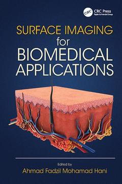
Based on hospital clinical trials examining the use of signal and image processing techniques, Surface Imaging for Biomedical Applications bridges the gap between engineers and clinicians. This text offers a thorough analysis of biomedical surface imaging to medical practitioners as it relates to the diagnosis, detection, and monitoring of skin conditions and disease. Written from an engineer?s perspective, the book discusses image acquisition methods, image processing, and pattern recognition techniques. It focuses on a variety of techniques used in recent years for image processing and pattern recognition (principal component analysis, independent component analysis, singular value decomposition, texture modeling, inverse model analysis, polynomial surface fitting, and classification techniques), and considers interventional and non-invasive procedures used to diagnose skin-related disease.
It examines the biological causation of four skin disorders (psoriasis, vitiligo, ulcer, and acne), provides basic terminologies in surface imaging, and details the outcome of various clinical observations and other research. It also details numerous measurement parameters related to surface imaging (body surface, skin color, tissue characteristic, thickness, roughness, volume of skin, and retinal changes).
- Discusses the development of a psoriasis severity measurement tool
- Provides material on assessing segmented repigmentation areas in vitiligo patients via VT-Scan
- Introduces a volume ulcer assessment using non-invasive 3D imaging
- Presents an automated system for acne grading that is based on capturing the images of various body parts using the DSLR camera
- Includes the MATLAB® codes for various pattern recognition techniques applied during the assessment/measurement at the end of each chapter
This interdisciplinary reference highlights the importance of disease diagnosis and monitoring, and is suitable for medical practitioners, biomedical engineers, and core image processing researchers.
Skin Surface Roughness Measurement for Assessing Scaliness of Psoriasis Lesions. Determination of Lesion Color for Clustering Psoriasis Erythema. Body Surface Area Measurement for Lesion Area Assessment. Skin Lesion Thickness Assessment. Analysis of Skin Pigmentation. Quantitative Assessment of Ulcer Wound Volume. Grading of Acne Vulgaris Lesions.
Ahmad Fadzil Mohamad Hani heads the Centre for Intelligent Signal & Imaging Research (CISIR), a university centre of excellence under the Mission-Oriented in Biomedical Engineering at Universiti Teknologi PETRONAS, Perak, Malaysia. He graduated with a BSc (1st Class Honors) in electronic engineering in 1983, obtained his MSc in telematics in 1984, and his PhD in image processing in 1991 from the University of Essex, UK. He is a Fellow of the Academy of Sciences Malaysia and a Fellow of the Institution of Engineers Malaysia. His current research challenges are developing new analysis techniques for early osteoarthritis and drug addiction using MRI techniques and bio-optics for skin pigmentation analysis. Professor Fadzil has authored over 190 research articles in journals and conferences proceedings, granted several patents, and won several awards for his work.
Date de parution : 04-2017
15.6x23.4 cm
Disponible chez l'éditeur (délai d'approvisionnement : 14 jours).
Prix indicatif 129,58 €
Ajouter au panierDate de parution : 06-2014
Ouvrage de 184 p.
15.6x23.4 cm
Disponible chez l'éditeur (délai d'approvisionnement : 15 jours).
Prix indicatif 232,80 €
Ajouter au panierThèmes de Surface Imaging for Biomedical Applications :
Mots-clés :
PASI Scoring; Membership Functions; Skin Surface Roughness Measurement for Assessing Scaliness of Psoriasis Lesions; Membership Degrees; Determination of Lesion Color for Clustering Psoriasis Erythema; Psoriasis Lesions; Body Surface Area Measurement for Lesion Area Assessment; Lesion Model; Analysis of Skin Pigmentation; Lesion Surface; Grading of Acne Vulgaris Lesions; Normal Skin; FCM Algorithm; FCM Cluster; Ul Ce; Gag; Vitiligo Lesion; Vitiligo Treatment; Acne Lesions; Acne; Midpoint Projection; Ulcer Wound; Lesion Thickness; RGB Image; CIELAB Color Space; Lesion Base; Lesion Area; Pe Rc; Fair Skin Tone; Convex Hull