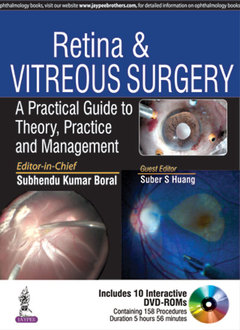
Retina & Vitreous Surgery A Practical Guide to Theory, Practice and Management
Auteurs : Boral Subhendu Kumar, Huang Suber S

This book is a comprehensive guide to retina and vitreous surgery for practising ophthalmic surgeons.
Each procedure is presented in a step by step format with discussion on theoretical background, retina pathophysiology, clinical signs and symptoms, investigations, differential diagnosis, and practical surgical steps with case discussions and numerous intraoperative photographs and surgical diagrams.
The book is accompanied by ten DVD ROMs demonstrating 158 surgical procedures, and edited by internationally recognised ophthalmic specialists.
Key Points
- Comprehensive guide to retina and vitreous surgery for ophthalmic surgeons
- Procedures presented in step by step format
- Book accompanied by ten DVD ROMs demonstrating 158 surgical procedures
- Highly illustrated with nearly 800 clinical photographs, diagrams and figures
- Chapter 1: Vitrectomy Machine, Cutter and Instrumentation
- Chapter 2: Visualization System
- Chapter 3: Illumination System
- Chapter 4: Cryoretinopexy and Endolaser Retinopexy
- Chapter 5: Intravitreal Tamponading Agents
- Chapter 6: Perfluorocarbon Liquids
- Chapter 7: Intravitreal Injections
- Chapter 8: Ophthalmic Ultrasonography
- Chapter 9: Steps of Making Sutureless Sclerotomy Wound
- Chapter 10: Vitrectomy
- Chapter 11: Diabetic Vitreous Surgery
- Chapter 12: Subretinal Hematoma with Vitreous Hemorrhage
- Chapter 13: Management of Retinal Detachment
- Chapter 14: Macular Surgeries
- Chapter 15: Managing Complications After Cataract Surgeries
- Chapter 16: Blunt Trauma, Penetrating Injuries and Retained Intraocular Foreign Bodies
- Chapter 17: Endophthalmitis
- Chapter 18: Combined Phacoemulsification and Vitreoretinal Intervention
- Chapter 19: 27-G Vitrectomy
- Chapter 20: Glued Intraocular Lens Surgeries
- Chapter 21: Intraocular Parasites
- Chapter 22: Silicone Oil Removal
- Chapter 23: Interface Vitrectomy
- Chapter 24: Managing Complications of Vitreoretinal Surgeries
- Chapter 25: How to Setup a Good Vitreoretinal Surgery Theater and Recording System?
DVD Content
- Chapter 3: Illumination System
- Video 1: Surgery with single-fiber endoillumination
- Video 2: Surgery with dual-fiber endoillumination
- Video 3: Surgery with Yellow filter
- Video 4: Surgery with Amber filter
- Chapter 4: Cryoretinopexy and Endolaser Retinopexy
- Video 1: Intraoperative cryo application
- Video 2: Endolaser application
- Chapter 6: Perfluorocarbon Liquids
- Video 1: Use of PFCL in RD
- Video 2: Use of PFCL in PVR
- Video 3: Use of PFCL in dropped nucleus—Animation
- Chapter 7: Intravitreal Injections
- Video 1: Intravitreal Triamcinolone Acetonide injection
- Video 2: Intravitreal anti-VEGF Injection
- Chapter 9: Steps of Making Sutureless Sclerotomy Wound
- Video 1: Port making in 23:25:27 gauze vitrectomy systems
- Chapter 10: Vitrectomy
- Video 1: Vitrectomy animation
- Video 2: Steps of vitrectomy in vitreous hemorrhage
- Video 3: Steps to manage retro lental or retro IOL blood
- Video 4: Steps to approach subhyaloid hemorrhage
- Chapter 11: Diabetic Vitreous Surgery
Part A: Management of Vitreomacular Interface Disorders
- Video 1: Identification of posterior hyaloid face—Application of triamcinolone acetonide
- Video 2: Removal of posterior hyaloid face by cutter (Animation + Video)
- Video 3: Identification and removal of vitreoschisis
- Video 4: Symptomatic vitreomacular adhesion (VMA)
- Video 5: Release of vitreomacular traction (VMT) by bent 24 G needle
- Video 6: Release of vitreomacular traction (VMT) by visco dissection
- Video 7: Vitreomacular traction with tractional retinal detachment
- Video 8: Epiretinal membrane
- Video 9: Refractory diabetic macular edema
- Video 10: Diabetic macular hole
Part B: Membrane Peeling
- Video 11: Forceps peeling—Animation + video
- Video 12: Chopstick peeling—Animation + video
- Video 13: Suction peeling—Animation + video
- Video 14: Segmentation—Animation + video
- Video 15: Delamination—Animation + video
- Video 16: Hydrodissection
- Video 17: Viscodissecton
Part C: Steps to Manage TRDs
- Video 18: Managing extramacular TRD
- Video 19: Managing macula-threatening TRD
- Video 20: Managing advanced TRDs
Part D: Steps to Manage Complex Combined Retinal Detachments
- Video 21: Localized macular combined RD
- Video 22: Managing complex combined RD
- Video 23: Bimanual vitrectomy in diabetics
- Chapter 12: Subretinal Hematoma with Vitreous Hemorrhage
- Video 1: Submacular hematoma-subretinal membrane removal
- Video 2: Submacular blood removal with full-thickness RPE-choroid graft transplantation
- Chapter 13: Management of Retinal Detachment
- Video 1: Scleral buckling (Nondrainage)
- Video 2: Scleral buckling with subretinal fluid drainage (Chandelier assisted)
- Video 3: General steps of vitreoretinal approaches to treat retinal detachment
- Video 4: Vitrectomy—Core and peripheral vitrectomy
- Video 5: PVD induction without PFCL—After core and peripheral vitrectomy
- Video 6: PVD induction with PFCL
- Video 7: PVD induction by forceps
- Video 8: Membrane peeling and sub-PFCL membrane peeling by forceps
- Video 9: Bimanual membrane peeling
- Video 10: Removal of subretinal band
- Video 11: Relaxing retinotomy for intrinsic retinal contraction
- Video 12: Extensive relaxing retinotomy for anterior PVR
- Video 13: Vitreous base excision with PFCL
- Video 14: Vitreous base excision without PFCL
- Video 15: Internal drainage of subretinal fluid by creating posterior retinotomy during FAE
- Video 16: Internal drainage of subretinal fluid through peripheral breaks using PFCL
- Video 17: Retinal detachment with dropped nucleus
- Video 18: Retinal detachment with dropped IOL
- Video 19: Retinal detachment with fundal coloboma with breaks at margin of coloboma
- Video 20: Retinal detachment with fundal coloboma with peripheral breaks
- Video 21: GRT with macula on retinal detachment
- Video 22: GRT with macula-off subtotal retinal detachment
- Video 23: Retinal detachment with GRT with inverted flap
- Video 24: Retinal detachment with GRT with rolled out margin
- Video 25: Retinal detachment with macular hole—ILM peeling on mobile retina
- Video 26: Retinal detachment with macular hole—ILM peeling under PFCL
- Video 27: Retinal detachment with choroidal detachment with suprachoroidal infusion
- Video 28: Retinal detachment with choroidal detachment
- Video 29: Retinal detachment with choroidal detachment with drainage of choroidal fluid
- Video 30 and 31: Bimanual surgeries for peripheral vitreous dissection and use two Chandelier light sources
- Video 32: Bimanual surgeries for removal of subretinal band
- Video 33: Bimanual surgeries to release fixed folds bimanually
- Chapter 14: Macular Surgeries
- Video 1: Epimacular membrane removal surgery
- Video 2: Vitrectomy with PVD induction by cutter in macular hole surgery
- Video 3: PVD induction by silicone tipped flute needle: Extrusion needle in macular hole surgery
- Video 4: Brilliant Blue G dye application under air or directly in water in macular hole surgery
- Video 5: Initial flap formation—By Tano scraper or end gripping forceps in macular hole surgery
- Video 6: ILM peeling in clockwise direction in macular hole surgery
- Video 7: ILM peeling in anticlockwise direction in macular hole surgery
- Video 8: Trypan Blue assisted ILM peeling in macular hole surgery
- Video 9: Inverse flap technique in large-sized macular hole surgery
- Video 10: Check vitreous base to rule out presence of any retinal break
- Video 11: Fluid-air exchange
- Video 12: Surgery for traumatic macular hole
- Video 13: Surgery for idiopathic partial thickness macular hole
- Video 14: Surgery for myopic foveoschisis
- Video 15: Surgery for optic disc pit with maculopathy
- Chapter 15: Managing Complications after Cataract Surgeries
- Video 1: Dropped cortex
- Video 2: Dropped epinuclear fragments
- Video 3: Dropped nucleus
- Video 4: Dropped IOL removal by end gripping forceps
- Video 5: Dropped IOL removal with PFCL
- Video 6: Dropped PCIOL removal by passive suction
- Video 7: Dropped PCIOL removal by active suction
- Video 8: Suprachoroidal hemorrhage management—Animation
- Video 9: Suprachoroidal hemorrhage management—Case I
- Video 10: Supra choroidal hemorrhage management—Case II
- Chapter 16: Penetrating Injuries and Retained Intraocular Foreign Bodies
- Video 1: Metallic foreign body with clear lens
- Video 2: Metallic foreign body with cataract
- Video 3: Metallic foreign body with endophthalmitis
- Video 4: Nonmetallic foreign body
- Chapter 17: Endophthalmitis
- Video 1: Animation
- Video 2: Intravitreal antibiotic injection—Technique
- Video 3: Surgical management of postoperative endophthalmitis
- Chapter 18: Combined Phacoemulsification and Vitreoretinal Interventions
- Video 1: Animation
- Video 2: Combined surgery for cataract with macular surgery
- Video 3: Combined surgery for cataract with vitreous hemorrhage
- Video 4: Combined surgery for cataract with retinal detachment
- Chapter 19: 27G Vitrectomy
- Video 1: Surgery for vitreous hemorrhage
- Video 2: Surgery for epiretinal membrane
- Video 3: Surgery for macular holes
- Video 4: Surgery for retinal detachment
- Video 5: Surgery for complex diabetic detachment
- Chapter 20: Glued IOL Surgeries
- Video 1: Standard Glued IOL surgery
- Video 2: Managing dropped scleral fixated IOL
- Video 3: Dislocation of IOL-CTR complex
- Chapter 21: Intraocular Parasites
- Video 1: Intraocular Gnathostomata with retinal detachment
- Video 2: Intraocular Cysticercosis with retinal detachment
- Video 3: Intraocular Toxocariasis with midperipheral granuloma
- Video 4: Intraocular Toxocariasis with inferior vitreous band and macular tractional fold
- Chapter 22: Silicone Oil Removal
- Video 1: Silicone oil removal passively
- Video 2: Silicone oil removal by active suction
- Video 3: Advanced emulsification of silicone oil
- Video 4: Combined phacoemulsification and silicone oil removal
- Chapter 23: Interface Vitrectomy
- Video 1: Interface vitrectomy under air
- Video 2: Interface vitrectomy at the margin of PFCL
- Video 3: Interface vitrectomy under silicone oil
- Chapter 24: Managing Complications of Vitreoretinal Surgeries
Part A: Preoperative Complications
- Video 1: Standard methods of ocular anesthesia and needle perforation with (Animation and Video)
- Video 2: Perforation with vitreous hemorrhage (Animation and Video)
- Video 3: Perforation with vitreous hemorrhage with partial retinal detachment
- Video 4: Perforation with vitreous hemorrhage with total retinal detachment (Animation and Video)
- Video 5: Perforation with macular break with retinal detachment (Animation and Video)
- Video 6: Perforation with hemorrhagic retinal detachment with retinal incarceration
Part B: Intraoperative Complications
- Video 7: Cathotomy for small palpebral fissure
- Video 8: Slippage of infusion cannula
- Video 9: Suprachoroidal infusion
- Video 10: Subretinal infusion
- Video 11: Retinal incarceration at the cannula tip
- Video 12: Complications during phaco-vitrectomy—Iris prolapse during dye injection
- Video 13: Complications during phaco-vitrectomy—Sudden influx of air bubbles
- Video 14: Complications during PVD induction with attached retina
- Video 15: Complications during PVD induction with detached retina—Inadvertent break
- Video 16: Intraoperative sudden unplanned hypotony
- Video 17: Intraoperative media haziness
- Video 18: Stepwise approach to manage intraocular bleed—Cautery
- Video 19: Stepwise approach to manage intraocular bleed—Raising IOP
- Video 20: Stepwise approach to manage intraocular bleed—Start FAX in high IOP
- Video 21: Stepwise approach to manage intraocular bleed—Application of triamcinolone acetonide over the bleeders
- Video 22: Using heavy fluid PFCL over the posterior pole
- Video 23: Intraoperative retinal break with retinal detachment
- Video 24: Subretinal migration of PFCL bubbles
Part C: Postoperative Complications
- Video 25: Phacoemulsification for postvitrectomy cataract
- Video 26: Submacular PFCL removal
- Video 27: Failed retinal detachment—Case of missed break
- Video 28: Failed retinal detachment—Case of ERM with new break
- Video 29: Failed retinal detachment—Case of ERM with reopening of break
- Video 30: Failed retinal detachment—Case with reopening of inferior break with inferior fixed fold with subretinal band
Subhendu Kumar Boral MD (AIIMS) DNB
Senior Vitreoretina Consultant, Disha Eye Hospitals, Kolkata, India
Suber S Huang MD MBA
CEO, Retina Centre of Ohio, Cleveland, Ohio; Chair, National Eye Health Education Committee, National Eye Institute, National Institutes of Health, Bethesda, USA
Date de parution : 10-2016
Ouvrage de 246 p.
21.5x27.9 cm
Disponible chez l'éditeur (délai d'approvisionnement : 14 jours).
Prix indicatif 428,93 €
Ajouter au panier


