Musculoskeletal Sonography, 2007 Technique, Anatomy, Semeiotics and Pathological Findings in Rheumatic Diseases
Coordonnateurs : Martino Fabio, Silvestri Enzo, Grassi Walter, Garlaschi Giacomo
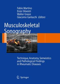
This book elucidates on the examination technique, the sonographic changes in musculoskeletal rheumatic involvement and the ultrasound assessment of joint rheumatic diseases. The atlas is enriched with several figures, in which the US picture is compared with that of conventional radiography, CT and MRI. It provides a unique collection of black and white and color images for easy and reliable diagnosis. The book is a practice-oriented tool.
Equipment and examination technique generalities. Equipment. Examination technique. Artifacts. Doppler technologies. Echo-contrast agents.- Technical examination procedure. Thoracic and abdominal wall. Upper extremity. Lower extremity.- Sonographic and power Doppler normal anatomy. Cartilage. Synovial spaces. Tendons and ligaments. Muscles. Nerves. Dermis and hypodermis.- Sonographic changes in musculoskeletal rheumatic involvement. Cartilage. Synovial spaces. Tendons and ligaments. Muscles. Nerves. Dermis and hypodermis.- Pathologic findings in rheumatic diseases. Osteoarthritis. Rheumatoid arthritis. Seronegative spondyloarthritis. Crystal-related arthropathies. Connective tissue disorders. Metabolic diseases. Synovial osteochondromatosis. Pigmented villonodular synovitis. Septic arthritis. Hemophiliac arthropathy. Primitive and peripheral entrapment neuropathies. Rheumatology in sport.- Ultrasonography and therapy monitoring.- Ultrasound-guided procedures.
Unique collection of black and white and color images for easy and reliable diagnosis
Practice-oriented, invaluable tool for professionals and residents
Most didactic approach supported by a specific page layout and graphic
Date de parution : 11-2006
Ouvrage de 207 p.
24.2x31.2 cm