L’édition demandée n’est plus disponible, nous vous proposons la dernière édition.
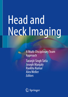
Head and Neck Imaging, 1st ed. 2021 A Multi-Disciplinary Team Approach
Langue : Anglais

This book provides a practically applicable guide to the all the different imaging modalities used in the diagnosis and management of ENT & Head and Neck patients. It bridges the gap in understanding between surgeons treating ENT & Head and Neck conditions and radiologists who oversee the process of scan requests, interpretation and delivering reports that best inform the subsequent management. Chapters cover a variety of sub-specialist areas including plain films, ultrasound, computed tomography (CT), magnetic resonance imaging (MRI), auditory implantation, paediatrics, head and neck cancer, trauma, three dimensional (3D) reconstruction and rehabilitation including swallow. This book facilitates surgeons and radiologists to further develop their understanding of each other?s perspectives on clinical decision-making and appropriately interpreting the outputs from a range of imaging modalities.
Head and Neck Imaging: A Multi-Disciplinary Team Approachis a resource well-suited to all trainees, residents, consultants who use these techniques to treat patients with head and neck symptoms. Furthermore, it is vital for those individuals preparing for exams in disciplines such as ear nose and throat, maxillofacial surgery and radiology.
On Call Modality Selection: Is the plain film dead?.- Head & Neck Ultrasound for Acute Admissions and in the Lump Clinic.- CT Workhorse.- Head and Neck Fascial Planes and Deep Neck Space Imaging.- Imaging the Unified Airway.- Friday Night Head and Neck Trauma.- Imaging Of The Temporal Bone in Hearing Loss.- Radiology of Head and Neck Cancer.- Sino-nasal Radiology.- Benign salivary gland disease: Imaging , diagnosis and minimal invasive treatment.- Radiological imaging for non-traumatic paediatric ENT conditions.- Evidence Based Imaging for Thyroid and Parathyroid Disease Management.- Imaging of the Lateral Skull Base and Cochlear Implants.- Anterior skull base and sinonasal surgery: dilemmas and complexities in management.- Imaging of Swallow.- The problematic middle ear and Cholesteatoma.- Challenges in Sinonasal & Anterior Skull Base Imaging.- Dysphagia following treatment for head and neck cancer.- Imaging Considerations for Laryngeal Cancer Surgery.- 3D Imaging, 3D Printing and Additive Manufacture in Complex Reconstruction & Craniofacial Surgery Planning.- Imaging for Anterior Neck Trauma.
Taranjit Singh Tatla
Taran Tatla is Training Program Director for ENT Higher Surgical Training, London North Thames and a Consultant ENT-Head & Neck-Thyroid Surgeon based at London North West University Healthcare NHS Trust. Achieving First Class Intercalated BSc Honours in Anatomy and Developmental Biology, he graduated with MBBS from University College London. Postgraduate basic and higher surgical training incorporated posts at many of the renowned London teaching hospitals with FRCS (ORL-HNS) award from Royal College of Surgeons, England. He completed a PhD in Applied Optical Imaging linked with the Hamlyn Centre for Robotics and Department for Surgery and Cancer, Imperial College London. He runs a number of innovative, team-focused multi-disciplinary postgraduate courses and serves as NIHR ENT Clinical Research Lead for North West London. He serves as a council member for the British Laryngological Association and Honorary Secretary for ENT UK.
Joseph Manjaly
Joseph Manjaly is a Consultant ENT Surgeon at the Royal National ENT Hospital and University College London Hospitals, specialising in Otology and Auditory Implant surgery. He completed higher surgical training in the London North Thames region and subsequently undertook a fellowship in otology and hearing implantation at Cambridge University Hospitals. He has held an interest in teaching since his undergraduate years at Bristol University and has co-authored a number of trainee textbooks widely used in the UK and abroad. He has been actively involved in training issues regionally and nationally, holding a number of committee roles. As well as being a keen sports fan he is also a musician who performs in a band semi-professionally around the country.
Raekha Kumar
Raekha Kumar is a Consultant Radiologist at West Hertfordshire Hospitals NHS Trust, specialising in Head and Neck Radiology. She graduated from
This book covers a full spectrum of topics spanning from basic principles to complex considerations The book is fully illustrated and incorporates schematic diagrams, tables and charts, making it more reader-friendly The book includes correlations of 2-D and 3-D anatomy teaching through the use of developing technology and software solutions, allowing new innovative methods of anatomy illustrations to be presented
Date de parution : 11-2021
Ouvrage de 459 p.
17.8x25.4 cm
Thèmes de Head and Neck Imaging :
© 2024 LAVOISIER S.A.S.
Ces ouvrages sont susceptibles de vous intéresser

Introductory Head & Neck Imaging 52,23 €


