From Signals to Image, 1st ed. 2020 A Basic Course on Medical Imaging for Engineers
Langue : Anglais
Auteurs : Azhari Haim, Kennedy John A., Weiss Noam, Volokh Lana
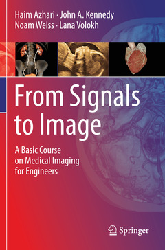
This textbook, intended for advanced undergraduate and graduate students, is an introduction to the physical and mathematical principles used in clinical medical imaging. The first two chapters introduce basic concepts and useful terms used in medical imaging and the tools implemented in image reconstruction, while the following chapters cover an array of topics such as physics of x-rays and their implementation in planar and computed tomography (CT) imaging; nuclear medicine imaging and the methods of forming functional planar and single photon emission computed tomography (SPECT) images and Clinical imaging using positron emitters as radiotracers. The book also discusses the principles of MRI pulse sequencing and signal generation, gradient fields, and the methodologies implemented for image formation, form flow imaging and magnetic resonance angiography and the basic physics of acoustic waves, the different acquisition modes used in medical ultrasound, and the methodologies implemented for image formation and flow imaging using the Doppler Effect.
By the end of the book, readers will know what is expected from a medical image, will comprehend the issues involved in producing and assessing the quality of a medical image, will be able to conceptually implement this knowledge in the development of a new imaging modality, and will be able to write basic algorithms for image reconstruction. Knowledge of calculus, linear algebra, regular and partial differential equations, and a familiarity with the Fourier transform and it applications is expected, along with fluency with computer programming. The book contains exercises, homework problems, and sample exam questions that are exemplary of the main concepts and formulae students would encounter in a clinical setting.
Introduction.- Basic Principles of Image Reconstruction.- X-ray Imaging and Computed Tomography.- Nuclear Medicine: Planar and SPECT Imaging.- Positron Emission Tomography (PET).- Magnetic Resonance Imaging (MRI).- Ultrasound Imaging.- Hybrid and Novel Methods.- Appendix.- Exemplary Solved Problems and Exams.
Haim Azhari is an Associate Professor in the Department of Biomedical Engineering, Technion, Israel Institute of Technology in Haifa, Israel.
John A. Kennedy is Chief Physicist in the Department of Nuclear Medicine, Rambam Health Care Campus in Haifa, Israel and is Adjunct Lecturer, Faculty of Biomedical Engineering, Technion, Israel Institute of Technology.
Lana Volokh is a medical imaging and image reconstruction professional, and an expert in nuclear medicine and hybrid imaging systems and image quality assessment.
Noam Weiss, PhD, is a medical physicist researcher and an expert in X-Ray CT technology and medical image processing. She is also an adjunct lecturer at the Faculty of Biomedical Engineering, Technion, Israel Institute of Technology.
Teaches the basic principles of medical imaging needed for understanding and conducting research and development in the field Emphasizes practical calculations for the design and evaluation of medical imaging devices Contains exemplary exercises, homework problems, and sample exam questions
Date de parution : 05-2021
Ouvrage de 474 p.
15.5x23.5 cm
Date de parution : 05-2020
Ouvrage de 474 p.
15.5x23.5 cm
Thèmes de From Signals to Image :
Mots-clés :
RADON transform; Medical imaging textbook; Analytical reconstruction; Algebraic reconstruction tomography; X-ray and computed tomography; Planar imaging; Dosimetry and safety; Radioisotopes and radioactive decay; Signal sources; Positron emission tomography (PET); Magnetic resonance imaging (MRI); Ultrasound imaging; Doppler shift; Acoustic impedance; Limited angle tomography; Compressed sensing; Basic concepts in signal processing; Nuclear medicine
© 2024 LAVOISIER S.A.S.