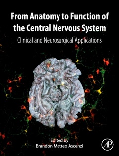
From Anatomy to Function of the Central Nervous System Clinical and Neurosurgical Applications
Coordonnateur : Ascenzi Brandon Matteo
1. Development of the nervous system and human neural stem cells
2. Topography of neuraxis, vascularization and ventricular system
3. Neurochemistry of the central nervous system
Section II: Anatomy of the spinal cord and brain
4. The spinal cord
5. The brainstem and reticular formation
6. The cerebellum
7. The diencephalon: introduction and epithalamus
8. The diencephalon: dorsal thalamus
9. The diencephalon: subthalamus or ventral thalamus
10. The diencephalon: hypothalamus
11. The telencephalon: introduction and cerebral cortex
12. The telencephalon: cingulate cortex
13. The telencephalon: hippocampus and related structures
14. The telencephalon: amygdala and claustrum
15. The telencephalon: basal ganglia
16. Circumventricular organs
Section III: Functional systems
17. General sensory systems and taste
18. The vestibular system
19. The auditory system
20. The visual system
21. Motor systems
22. The limbic system
Section IV: Appendix
Dr Ascenzi received his Bachelor’s Degree in Techniques of Biomedical Laboratory and his Master’s Degree in Neurobiology (Medical Biotechnology) at School of Medicine and Surgery, University of Rome “Tor Vergata. Over the years he took part in several research project about neuromuscular diseases, such as Duchenne’s Muscular Dystrophy, at the Institute of Histology and Embryology of University of Rome “La Sapienza and at the School of Sciences (MM.PH.NN) of “Tor Vergata University.
Since 2015 to middle 2016 he worked, at National Research Council “CNR, to a project on the mechanism of maturation and secretion of Nerve Growth Factor (NGF) in a murine model of diabetic and Alzheimer-like encephalopathy. In 2017 he won the European PhD “Marie Curie fellowship and he worked at the Centro de Biologia Molecular “Severo Ochoa of Madrid in collaboration with University of Lund, Polytechnic University of Milan, Barcelona, Oslo, Munich and others. His fields of research interest mainly focus on the modulation of brain development through neurotransmitters and peptides in the field of neurodevelopmental diseases (ASD, ADHD). Recently he also started to participate at neurosurgical and radiological works. Currently he is majoring in Medicine, he is 33 years old and this is his first monography.
- Integrates anatomo-functional descriptions with the synaptic organization of the CNS
- Allows readers to better understand the morphology and topography of encephalic structures
- Features neuroradiological images and drawings of brains dissected with Klingler’s method
- Chapters have references (key articles, books, protocols) for additional detailed study
Date de parution : 09-2024
Ouvrage de 800 p.
21.4x27.6 cm