Dermatoscopy of Non-Pigmented Skin Tumors Pink - Think - Blink
Coordonnateurs : Zalaudek Iris, Argenziano Giuseppe, Giacomel Jason
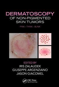
Although many skin lesions are pigmented, Dermatoscopy of Non-pigmented Skin Tumors: Pink - Think - Blink addresses non-pigmented lesions, which may be more difficult to diagnose. It discusses dermatoscopy not only as a reliable tool for diagnosis, but also for the monitoring of treatment outcomes following topical therapy.
The clinical diagnosis of non-pigmented skin lesions is one of the most challenging in the daily routine. To arrive at a correct diagnosis?or at least an adequate management plan?the clinician needs to collect many pieces of information and put them together like a puzzle. Illustrated with nearly 200 color clinical and dermatoscopic photographs, this book is an invaluable guide for clinicians striving to solve the diagnostic puzzle and correctly identify non-pigmented lesions.
TECHNICAL ASPECTS. Physics of Polarized and Nonpolarized Dermoscopy and Digital Photography. Instrument-Dependent Criteria. Metaphoric and Descriptive Language in Dermoscopy: Lessons fromthe Cognitive Sciences.How to Perform Dermoscopy of Non-Pigmented Skin Lesions. DERMATOSCOPY OF NON-PIGMENTED LESIONS. How to Assess a Given Non-Pigmented Lesion. Clinical Assessment. Vascular Morphologies. Vascular Arrangements. Specific Patterns. Dermatoscopic Clues in Non-Pigmented Lesions. The Influence of Tumor Thickness on the Vascular Morphologies. SPECIFIC DERMATOSCOPIC PATTERNS AND THEIR DIAGNOSTIC SIGNIFICANCE. Intradermal Nevi (Including Unna and Miescher Type). Clark Nevi in Fair Skin Types. Spitz Nevi. Atypical Spitzoid Neoplasms (Atypical Spitz Nevi, Atypical Spitz Tumors, Spitzoid Melanoma): a Clinicopathological Update. Nevi in Patients With Bap1 Germ Line Mutation, Red-Hair Polymorphism, and Albinism. Amelanotic Melanoma. Hypomelanotic Melanoma. Cutaneous Melanoma Metastases. Sebaceous Hyperplasia. Seborrheic Keratosis. Dermatofibromas. Angioma, Pyogenic Granuloma, Angiokeratoma. Benign Adnexal Lesions. Basal Cell Carcinoma. Keratinocyte Skin Cancer. Dermoscopy of Cutaneous Neuroendocrine ("Merkel cell") Carcinoma. Malignant Vascular, Adnexal, and Fibrous Tissue Tumors. Clues for the Differential Diagnosis of Inflammatory Lesions from Tumoral Lesions. Dermoscopy for Assessing Surgical Margins. Dermoscopy in the Treatment Decision (Surgical vs. Topical). Dermoscopy for Treatment Monitoring (Recurrence vs. Clearance). Diagnostic Clues and Management Rules. Confocal Microscopy in the Diagnosis and Management of Non-Pigmented Skin Tumors (Which, When, and When Not).
Edited by:
Iris Zalaudek, MD, Division of Dermatology and Venerology, Medical University of Graz, Austria
Giuseppe Argenziano, MD, Skin Cancer Unit, Arcispedale S. Maria Nuova IRCCS Reggio Emilia, Italy
Jason Giacomel, MBBS, Skin Spectrum Medical Services, Perth, Australia
Date de parution : 08-2015
17.8x25.4 cm
Thèmes de Dermatoscopy of Non-Pigmented Skin Tumors :
Mots-clés :
Spitz Nevi; Melanocytic Neoplasm; Dermatoscopy; Superficial Basal Cell Carcinoma; Dermoscopy; Clear Cell Acanthoma; Nonpigmented lesions; Seborrheic Keratoses; Dermatoscopy of nonpigmented lesions; Intradermal Nevi; Dermatoscopic patterns; Vice Versa; Non-pigmented lesions; Linear Irregular Vessels; Blue Gray Ovoid Nests; Dermoscopic Images; Dotted Vessels; Dermoscopic Feature; Pyogenic Granuloma; Structureless White Areas; Basal Cell Carcinoma; Hairpin Vessels; Amelanotic Melanoma; Coiled Vessels; Polymorphous Vessels; Nodular BCC; Comma Vessels; Nodular Melanoma; Hypomelanotic Melanoma; Sebaceous Hyperplasia