Dermatopathology of Tropical Diseases Pathology and Clinical Correlations
Auteurs : Sangüeza Omar P, Sangüeza Martin, Bravo Francisco
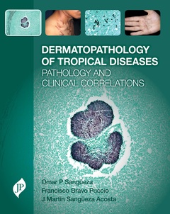
The majority of tropical diseases manifest themselves through the skin, and while many can be diagnosed on physical examination, dermatopathologic results are often of critical importance to the diagnostic process. However, general dermatopathology texts lack images of diseases endemic in the developing world; they also lack clinical correlates linking the pathology to the clinical entity. This is a problem, because greater movement between countries where tropical diseases are prevalent and Europe, North America and other developed nations makes it essential for physicians to be able to recognize, diagnose and treat these diseases.
Dermatopathology of Tropical Diseases covers the pathology and clinical correlations of the most important tropical diseases. Each entity is described using brief text that summarises its epidemiology, pathogenesis and clinical features. The diagnostic process is then described and illustrated using both clinical and histopathologic images. Wherever possible, different examples are shown of the same disease, in order to illustrate as wide a variety of presentations as possible.
Extensively illustrated with almost 600 photographs, Dermatopathology of Tropical Diseases is an invaluable resource for dermatopathologists, pathologists and dermatologists based in the developing world as well as those in developed countries who see tropical diseases in returned travellers and immigrant populations.
Key features
- Includes chapters on all major tropical diseases, with both clinical and histopathological images provided for each entity, allowing the reader to correlate pathological characteristics with the clinical presentation of each disease
- Wherever possible, different examples of the same disease are shown, to illustrate the variety of presentations
- Lavishly illustrated with almost 600 colour photographs
PART 1: BACKGROUND
- 1 Introduction
PART 2: VIRAL DISEASES
- 2 Orf and milker's nodules
- 3 Dengue
- 4 Hydroa vacciniforme and hydroa-like lymphomas
- 5 Infective dermatitis: a cutaneous manifestation of HTLV-1 infection
PART 3: BACTERIAL AND RICKETTSIAL INFECTIONS
- 6 Rhinoscleroma
- 7 Endemic treponematoses
- 8 Carrion’s disease
- 9 Plague
- 10 Cutaneous anthrax
- 11 Bacterial mycetomas: actinomycosis, botryomycosis, and nocardiosis
- 12 Rickettsiosis
- 13 Granuloma inguinale
- 14 Pitted keratolysis
- 15 Erythrasma
- 16 Tularemia
- 17 Tropical ulcer
PART 4: MYCOBACTERIAL INFECTIONS
- 18 Leprosy
- 19 Cutaneous tuberculosis
- 20 Buruli ulcer
- 21 Nontuberculous mycobacterial skin infections
PART 5: MYCOTIC INFECTIONS
- 22 Eumycotic mycetomas
- 23 Lobomycosis
- 24 Chromoblastomycosis
- 25 Tinea nigra
- 26 Phaeohyphomycosis
- 27 Sporotrichosis
- 28 Alternariosis
- 29 Hyalohyphomycoses
- 30 Zygomycosis
- 31 Paracoccidioidomycosis
- 32 Histoplasmosis
- 33 Rhinosporidiosis
- 34 Cryptococcosis
- 35 North American blastomycosis
PART 6: ARTHROPOD-INDUCED DISEASES
- 36 Venomous arthropods
- 37 Myiasis
- 38 Tungiasis
PART 7: PROTOZOAL INFECTIONS
- 39 Cutaneous leishmaniasis
- 40 Diseases caused by ameba
- 41 Chagas’ disease
PART 8: ALGAE INFECTIONS
- 42 Protothecosis
PART 9: HELMINTH INFESATIONS
- 43 Schistosomiasis
- 44 Cutaneous larva migrans
- 45 Gnathostomiasis
- 46 Cysticercosis
- 47 Sparganosis
- 48 Echinococcosis
- 49 Human filariasis
Omar P Sangüeza MD
Professor and Director of Dermatopathology, Wake Forest University School of Medicine, Winston-Salem, NC, USA
Martin Sangüeza MD
Dermatopathologist, Hospital Obrero Caja Nacional de Salud, Department of Pathology, La Paz, Bolivia
Francisco Bravo MD
Alexander von Humboldt Institute of Tropical Medicine, Universidad Peruana Cayetano Heredia, Lima, Peru
Date de parution : 03-2017
Ouvrage de 344 p.
21.9x27.5 cm
Disponible chez l'éditeur (délai d'approvisionnement : 14 jours).
Prix indicatif 177,01 €
Ajouter au panier