Congenital Heart Defects. Decision Making for Surgery, Softcover reprint of the original 1st ed. 2009 Volume 3: CT-Scan and MRI
Langue : Anglais
Auteurs : Corno Antonio F., Festa Pierluigi
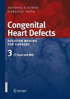
The diagnosis and management of congenital heart defects has rapidly evolved over the last few decades. In this third volume of the series entitled "Congenital Heart Defects: Decision Making for Surgery" Antonio Corno provides an up-to-date and comprehensive presentation of the new role that cardiac CT and MRI will play in the management of congenital heart defects. He has been ably assisted by a cardiologist, Pierluigi Festa. The book provides a dazzling array of images derived by both techniques and covers the full range of congenital heart malformations. Both the pre-operative and post-operative usefulness of these techniques is presented: in the pre-operative period with regard to the details useful for choosing among all available surgical options, in the post-operative period for monitoring the follow-up and potential complications. There is no doubt that these techniques will be particularly helpful for older children and adults with congenital heart disease in assessing the late impact of a congenital heart malformation and the surgical repair or palliation which may have been undertaken years previously. The time is definitely right for a comprehensive presentation of these new diagnostic modalities and the way in which they will influence the field of congenital heart management. First and only volume on the market presenting all the pre- and post-operative aspects and images of congenital heart defects as seen by cardiac CT and MRI. Numerous illustrations provide clear and educationally useful images for each specific anatomic detail. Helps to decide on the best surgical option. Describes potential complications. Provides advice with regard to monitoring in the post-operative period.
to CT scan and MRI.- Anomalous systemic venous connections.- Anomalous pulmonary venous connections.- Atrial septal defect.- Unroofed coronary sinus.- Atrioventricular septal defect.- Ventricular septal defect.- Tetralogy of Fallot.- Tetralogy of Fallot with absent pulmonary valve.- Pulmonary atresia with intact ventricular septum.- Ebstein’s anomaly.- Patent ductus arteriosus.- Left ventricular outflow tract obstruction.- Aortic coarctation.- Aortic arch interruption.- Complete transposition of the great arteries.- Hypoplastic left heart syndrome.- Cor triatriatrum.- Tricuspid atresia.- Single ventricle.- Pulmonary atresia with ventricular septal defect.- Truncus arteriosus.- Aortopulmonary window.- Anomalous pulmonary arteries.- Anomalous coronary arteries.- Double outlet right ventricle.- 3.26 Double discordance.- Isomerism.- Slings and rings.
First and only volume on the market presenting all the pre- and post-operative aspects and images of congenital heart defects as seen by CT and MRI Numerous illustrations provide clear and didactic images for each specific anatomic detail Helps to decide on the best surgical option Provides advice with regard to monitoring in the post-operative period Alerts to potential complications Includes supplementary material: sn.pub/extras
Ouvrage de 223 p.
21x27.9 cm
Thèmes de Congenital Heart Defects. Decision Making for Surgery :
© 2024 LAVOISIER S.A.S.