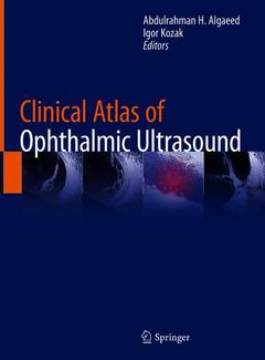
Clinical Atlas of Ophthalmic Ultrasound, 1st ed. 2019
Coordonnateurs : Algaeed Abdulrahman H., Kozak Igor

There have been significant advancements in the field of ophthalmic ultrasound as this imaging technology can now detect and differentiate minute lesions in a wide variety of eye disorders. With understanding of the indications for ultrasonography and proper examination techniques, one can gather a vast amount of information not possible with a clinical exam alone. Clinical Atlas of Ophthalmic Ultrasound includes a short clinical description of each case presented and supplemented with high quality, color fundus images, wide-field images, CT/MRI scans, and/or pathologic slides where applicable.
Written for ophthalmologists, radiologists, echographers, and ophthalmic oncologists, this book offers more of a comprehensive clinical view on a particular disease, including multimodal imaging approach, rather than just ultrasound characteristics. Chapters covering clinical and surgical globe anatomy, vitreo-retinal disease, trauma, intraocular tumors, and optic nerve disorders are all included.
1 History and Principles of Ocular Ultrasonography
2 Clinical Globe Anatomy
3 Vitreous/Retina/Choroid
4 Ocular Trauma/Endophthalmitis
5 Ocular Tumors
6 Optic Nerve
7 Sclera/Ciliary Body/Anterior Segment
8 Miscellaneous Case
Includes a short clinical description of each case along with high quality, color fundus images, wide-field images, CT/MRI scans, and/or pathologic slides where applicable
Written for ophthalmologists, radiologists, echographers, and ophthalmic oncologists
Offers more of a comprehensive clinical view on a particular disease, rather than just ultrasound characteristics
Chapters include clinical and surgical globe anatomy, vitreo-retinal disease, trauma, intraocular tumors, and optic nerve disorders
Date de parution : 02-2019
Ouvrage de 69 p.
21x27.9 cm


