Atlas of Optical Coherence Tomography of Macular Diseases
Auteurs : Gupta Vishali, Gupta Amod, Dogra Mangat R.
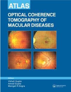
The emergence of Optical Coherence Tomography (OCT) in recent years has revolutionized the way we see the retina. Providing, in real time, high-resolution cross-sectional images of the macula that are very similar to obtaining in vivo histopathological specimens, OCT represents a major advance in the diagnostics of retinal disease. The excitement of working with this new tool has been dampened by the non-availability of any standard textbook on the subject and meant that every new finding on the OCT saw us rushing to the library almost on a daily basis to locate any published reports on the subject. Until now.
Containing nearly 900 scans of both normal and diseased appearances, most in full color, Atlas of Optical Coherence Tomography of Macular Diseases covers how to use Stratus OCT for diagnosing various macular disorders, identifying correct therapeutic approaches and monitoring the responses to therapies and interventions. The authors provide brief case summaries, fundus photographs, fluorescein angiography, and the OCT images and the follow up images. They discuss OCT applications for diagnosis, management, and follow-up in diabetic macular edema, macular hole, taut posterior hyaloid membrane, vitreofoveal traction, idiopathic central serous chorioretinoplasty, submacular pathology, and more.
Date de parution : 11-2004
Ouvrage de 320 p.
21x28 cm
Thèmes d’Atlas of Optical Coherence Tomography of Macular Diseases :
Mots-clés :
fluorescein; angiography; cystoid; edema; neurosensory; retina; retinal; pigment; epithelium; choroidal