Lavoisier S.A.S.
14 rue de Provigny
94236 Cachan cedex
FRANCE

Heures d'ouverture 08h30-12h30/13h30-17h30
Tél.: +33 (0)1 47 40 67 00
Fax: +33 (0)1 47 40 67 02


Url canonique : www.lavoisier.fr/livre/medecine/atlas-of-laser-scanning-ophthalmoscopy/descriptif_2288874
Url courte ou permalien : www.lavoisier.fr/livre/notice.asp?ouvrage=2288874

Atlas of Laser Scanning Ophthalmoscopy, 2004

Langue : Anglais

Auteurs :

Préfaciers : Völcker H.E., Pillunat L.E., Kruse F.E.
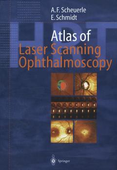
Couverture de l’ouvrage Atlas of Laser Scanning Ophthalmoscopy

This unique atlas is the most comprehensive and up-to-date reference of laser scanning ophthalmoscopy. It is ideal for residents and general ophthalmologists who want to enhance their diagnostic skills. The atlas contains superb images of all clinically relevant diseases diagnosed by current models of the Heidelberg Retina Tomograph. It correlates classical diagnostic tools such as perimetry, tonometry and fundus photography with state-of-the-art studies including digital retinal angiography, optical coherence tomography and laser scanning tomography. Special features include the illustrated coverage of diseases of the optic nerve head; different types and stages of glaucoma, and other topics.

1 Introduction.- 1.1 Why Is Laser Scanning Ophthalmoscopy Needed?.- 1.2 Technical Background.- 1.3 Stereometric Parameters of the HRT.- 2 Healthy Optic Discs.- 2.1 Natural Variety of Optic Discs.- 2.2 Regular Shapes.- 2.3 Micropapillas.- 2.4 Megalopapillas.- 2.5 Tilted Discs.- 3 Glaucomas.- 3.1 Automated Classification Procedures.- 3.2 Moderate Glaucomas.- 3.3 Nerve Fiber Bundle Defects.- 3.4 Advanced Glaucomas.- 3.5 Micropapillary Glaucomas.- 3.6 Megalopapillary Glaucomas.- 3.7 Normal Tension Glaucomas.- 3.8 Classification Errors.- 4 Follow-up Examinations.- 4.1 Strategies for Longitudinal Analysis.- 4.2 Progression of Glaucoma.- 5 Prominent Optic Discs.- 5.1 Scanning of Prominent Discs.- 5.2 Drusen of the Optic Nerve.- 5.3 Optic Nerve Swelling.- 5.4 Papilledemas.- 6 Macular Scans.- 6.1 The Macular Edema Software Module.- 6.2 Cystoid Macular Edema.- 6.3 Branch Vein Occlusion.- 6.4 Central Serous Retinopathy.- 6.5 Macular Puckers.- 6.6 Macular Holes.- 7 Miscellaneous.- 7.1 Optimizing Imaging.- 7.2 Astigmatism.- 7.3 Retinal Irregularities.- 7.4 Floaters.- 8 Confocal Corneal Imaging.- 8.1 The Rostock Cornea Modu.- References.

First atlas for interpreting Heidelberg Retina Tomograph pictures

Excellent teaching material

Includes supplementary material: sn.pub/extras

Date de parution :

Ouvrage de 170 p.

19.3x26 cm

Disponible chez l'éditeur (délai d'approvisionnement : 15 jours).

Prix indicatif 52,74 €

Ajouter au panier

Date de parution :

Ouvrage de 170 p.

20.3x27.6 cm

Sous réserve de disponibilité chez l'éditeur.

Prix indicatif 105,49 €

Ajouter au panier

Thème d’Atlas of Laser Scanning Ophthalmoscopy :