Atlas of Histology of the Juvenile Rat
Coordonnateurs : Parker George A, Picut Catherine A.
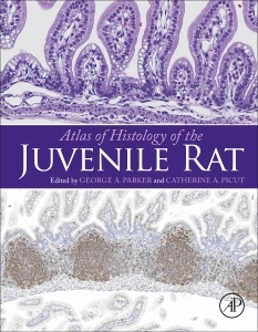
Atlas of Histology of the Juvenile Rat should be of interest to toxicologic pathologists, toxicologists, and other biological scientists who are interested in the histomorphology of juvenile rats. For several decades the laboratory rat has been used extensively in nonclinical toxicology studies designed to detect potential human toxicity of drugs, agrochemicals, industrial chemicals, and environmental hazards. These studies traditionally have involved young adult rats that are 8-10 weeks of age as studies are started. It is becoming increasingly apparent that children and young animals may have different responses to drug/chemical exposures, therefore, regulatory agencies are emphasizing toxicology studies in juvenile animals.
While the histologic features of organs from young adult and aged laboratory rats are well known, less is known about the histologic features of organs from juvenile rats. Final histologic maturity of many organs is achieved postnatally, thus immature histologic features must be distinguished from chemical- or drug-related effects. While this postnatal organ development is known to exist as a general concept, detailed information regarding postnatal histologic development is not readily available. The Atlas includes organs that are typically sampled in nonclinical toxicology studies and presents the histologic features at weekly intervals, starting at birth and extending through postnatal day 42.
Integumentary System, Including Mammary and Adnexal Glands
Lauren Staska
Musculoskeletal System
Jairo Nunes
Nervous System, Central and Peripheral
Amera Remick
Respiratory System
Melanie Greeley
Gastrointestinal System
Gary Coleman
Liver, Exocrine Pancreas and Salivary Glands
Danielle Brown
Reproductive System, Female
Catherine Picut
Reproductive System, Male
Catherine Picut
Endocrine System
Amera Remick
Immune System
George Parker
Hematopoietic System
Josely Figuieredo
Special Senses (Eye and Ear)
Gary Marit
Urinary System
Danielle Brown
Cardiovascular System
Melanie Greeley
Dr. Catherine Picut is a graduate of the University of Pennsylvania School of Veterinary Medicine, completed a residency program in veterinary pathology at Cornell University, and received a law degree from Yale University. She is a diplomate of the American College of Veterinary Pathologists and the American Board of Toxicology as well as a registered patent attorney and quality assurance professional (RQAP-GLP). She has 22 years of experience providing research pathology services on studies involving the safety evaluation of new drugs, biologics, medical devices and chemicals. She has published numerous articles in peer-reviewed scientific journals and has been author or co-author on numerous book chapters. Her special research interests are in reproductive and juvenile toxicology. Currently she is a senior pathologist at the Hillsboro
- Written and edited by highly experienced, board-certified toxicologic pathologists
- Includes more than 700 high-resolution microscopic images from organs that are typically examined in safety assessment toxicology studies
- Detailed figure legends and chapter narratives present the salient features of each organ at each time interval
- Figures are available for further study via Elsevier’s Virtual Microscope, which allows viewing of microscopic images at higher magnification
- Valuable resource for toxicologic pathologists who are confronted with interpretation of lesions in juvenile rats in situations where age-matched concurrent controls are not available for comparison, e.g., with unscheduled decedents
- Figures are available for further study on ScienceDirect with Virtual Microscope, which allows viewing of microscopic images at higher magnification
Date de parution : 05-2016
Ouvrage de 462 p.
21.4x27.6 cm