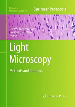
Light Microscopy, Softcover reprint of the original 1st ed. 2011 Methods and Protocols Methods in Molecular Biology Series, Vol. 689
Langue : Anglais

Of all scientific instruments, probably none has had more applications in the life sciences than the light microscope. In Light Microscopy: Methods and Protocols, expert researchers explore the basics and the latest advances in microscope instrumentation, sample preparation, and imaging techniques, all of which have been producing fundamental insights into the functions of cells and tissues. Chapters cover a variety of bright field and fluorescence microscopy-based approaches that are central to the study of a range of biological questions, providing information on how to prepare cells and tissues for microscopic investigations, covering detailed staining procedures, and exploring methods to analyze images and interpret the results accurately. Composed in the highly successful Methods in Molecular Biology? series format, each chapter contains a brief introduction, step-by-step methods, a list of necessary materials, and a Notes section which shares tips on troubleshooting and avoiding known pitfalls. Comprehensive and current, Light Microscopy: Methods and Protocols is an essential handbook for all researchers who are exploring the intriguing microscopic world of the cell.
Glycol methacrylate embedding for improved morphological, morphometrical and immunohistochemical investigations under light microscopy: testes as a model.- Histological processing of teeth and periodontal tissues for light microscopy analysis.- Large plant samples: how to process for 2-hydroxyethyl methacrylate embedding?.- Image cytometry: nuclear and chromosomal DNA quantification.- Histological approaches to study tissue parasitism during Trypanosoma cruzi infection.- Intravital microscopy to study leukocyte recruitment in vivo.- Introduction to fluorescence microscopy.- Using the fluorescent styryl dye FM1-43 to visualize synaptic vesicles exocytosis and endocytosis in motor nerve terminals.- Imaging lipid bodies within leukocytes with different light microscopy techniques.- EicosaCell – An immunofluorescent-based assay to localize newly synthesized eicosanoid lipid mediators at intracellular sites.- Nestin-driven green fluorescent protein as an imaging marker for nascent blood vessels in mouse models of cancer.- Imaging calcium sparks in cardiac myocytes.- Light microscopy in aquatic ecology: methods for plankton communities studies.- Fluorescence immunohistochemistry in combination with differential interference contrast microscopy for studies of semi-ultrathin specimens of epoxy resin-embedding samples.
Readily reproducible protocols for optimal histological observations under bright-field microscopy Provides full description of plastic resin embedding methods for plant and animal research Presents advanced methods to study leukocyte biology and inflammatory responses Imaging techniques applied to cytogenetic, cancer, neurobiology, ion mobilization, and aquatic ecology studies Includes immunocytochemistry methods and a comprehensive overview of fluorescence microscopy Includes supplementary material: sn.pub/extras
Ouvrage de 244 p.
17.8x25.4 cm
Ouvrage de 244 p.
17.8x25.4 cm
Thème de Light Microscopy :
© 2024 LAVOISIER S.A.S.
