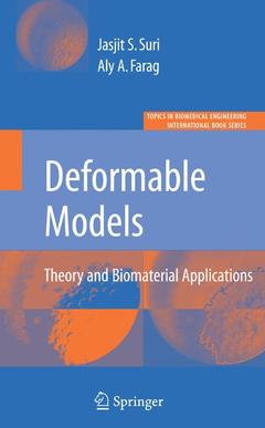
Deformable Models, 2007 Theory and Biomaterial Applications Topics in Biomedical Engineering. International Book Series
Coordonnateur : Farag Aly

In the biomedical field, biomedical imagery has come to be a discipline of its own, given the nature of its applications in the understanding of the human body and medical diagnostics. The understanding of Deformable Models are the significant utility on biomedical imagery primarily because of its ability to perform efficient topology preservation and fast shape recovery. This has dominated the binary, grayscale and color imaging frameworks, which the eye can perceive. It has not only the ability to find boundaries and surfaces that are deep-seated in 2-D and 3-D volumes respectively, but also provide satisfactory solutions for the completion of cognitive objects with missing boundaries.
Deformable Models: Theory and Biomaterial Applications focuses on the core image processing techniques: theory and biomaterials useful to research and industry.
Aly A. Farag received the bachelor degree from Cairo University, Egypt and the PhD degree from Purdue University in Electrical Engineering. He also holds master degrees in bioengineering from the Ohio State and the University of Michigan. He is a University Scholar and Professor of Electrical & Computer Engineering at the University of Louisville. Dr. Farag is the founder and director of the Computer Vision and Image Processing Laboratory (CVIP Lab) which focuses on imaging science, computer vision and biomedical imaging. Dr. Farag main research focus is 3D object reconstruction from multimodality imaging, and applications of statistical and variational methods for object segmentation and registration. He has authored over 250 technical papers in the field of image understanding and holds a number patents. He is regular reviewer to a number of professional organizations in the United States and abroad, and a member of the editorial boards of a number journals and international meetings.
Jasjit S. Suri, PhD is an innovator, scientist, a visionary, and industrialist and an internationally known world leader in Biomedical Engineering and Biological Sciences. Dr. Suri has spent over 20 years in the field of biomedical engineering, devices and its management. He received his Doctorate from the University of Washinton, Seattle and Master's in Executive Business Management from Weatherhead, Case Western Reserve University, Cleveland, Ohio. Dr. Suri was crowned with the President's Gold Medal in 1980 and elected as a Fellow of the American Institute for Medical and Biological Engineering.
Ouvrage de 581 p.
15.5x23.5 cm
Date de parution : 08-2007
Ouvrage de 584 p.
Thèmes de Deformable Models :
Mots-clés :
biomaterial; biomedical engineering; classification; diagnostics; entropy; image analysis; ultrasound


