Lavoisier S.A.S.
14 rue de Provigny
94236 Cachan cedex
FRANCE

Heures d'ouverture 08h30-12h30/13h30-17h30
Tél.: +33 (0)1 47 40 67 00
Fax: +33 (0)1 47 40 67 02


Url canonique : www.lavoisier.fr/livre/autre/cone-beam-ct-of-the-head-and-neck-an-anatomical-atlas/kau/descriptif_2324866
Url courte ou permalien : www.lavoisier.fr/livre/notice.asp?ouvrage=2324866

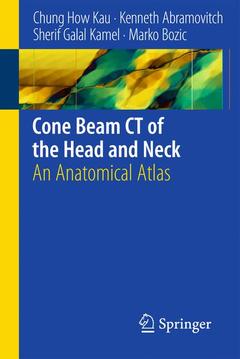
Cone Beam CT of the Head and Neck, 2011 An Anatomical Atlas

Langue : Anglais

Auteurs :

Couverture de l’ouvrage Cone Beam CT of the Head and Neck

'Cone Beam CT of the Head and Neck' presents normal anatomy of the head using photographs of cadavers and CBCT images in sagittal, axial and coronal planes with the anatomic structures and landmarks clearly labelled. Important structures and regions are presented in detailed view. The photographs of human tissue (based on slicing of cadaveric heads) combined with CBCT images have not been used previously for an atlas of anatomy. Scanned objects with the possibility of 3D reconstruction present better understanding of the anatomy.

Introduction.- Conventional Computed Tomography (CT).- CBCT.- CBCT Acquisition Systems.- Uses of CBCT Technology.- Axial.- Coronal.- Sagittal.
Professor Chung How KAU completed his dental training at the Faculty of Dentistry at the National University of Singapore after having received general practice in Cardiff, Wales. He actively contributes and publishes in the orthodontic literature with over 100 peer-reviewd publications, conference papers and lectures. In 2006, he was honored by the Hungarian Orthodontonic Society. Professor Kau is the winner of two prestigious research prizes of the British Orthodontic Society - the Chapman (2002) and Houston (2006) and received two research awards from the American Association of Orthodontists - the AAO Faculty Development Award (2006) and Southwestern Society of Orthodontist Research Award (2007). His research lab is in possession of US $ 250.000 worth of imaging equipment which includes the 3D Sirona Galileos cone beam scanner of the 3dMD surface imaging device. Professor KAU completed an AMBA accredited Business Program from the Leeds University Business School in 2003. He is a fully registered specialist and dentist in the United Kingdom and Singapore. He also participates in the Faculty Practice at the Dental Branch, Universiy of Texas Health Science Center.Marko BOZIC, MD: is a resident of maxillofacial surgery at the University Medical Centre in Ljubljana, Slovenia. He has finished the study of medicine in 2004 at the Medical Faculty in Ljubljana where he is currently finishing his Dental Medicine study and his PhD. Fulbright Scholar (UTHSC)Sherif Calal KAMEL, DDS MFDS RCSEdin received his education in the UK; International Research Scholarship (UTHSC)Kenneth ABRAMOVITCH; DDS MS is an Associate Professor and Head of Section (Oral Maxillofacial Radiology) at the Texas University, Houston, TX, USA.

Ultimate information on development from invention of X-Rays to CBCT (Cone Beam Computerized Tomograph Images)

Photos and CBCT images with clear anatomic labels represent basic learning and revision material First atlas with photographs of human tissue combined with CBCT images

Includes supplementary material: sn.pub/extras

Date de parution :

Ouvrage de 66 p.

Disponible chez l'éditeur (délai d'approvisionnement : 15 jours).

105,49 €

Ajouter au panier

Ces ouvrages sont susceptibles de vous intéresser