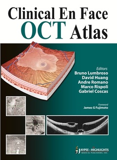
Clinical En Face OCT Atlas
Auteurs : Lumbroso Bruno, Huang David, Romano Andre, Rispoli Marco, Coscas Gabriel

This atlas examines developments in clinical en face imaging, comparing methods and devices and evaluating the most clinically efficient techniques. Divided into three sections, the first part introduces the principles of OCT (optical coherence tomography) and the anatomy and histology of the retina and surrounding area.
The second section discusses en face OCT in diagnosing and treating different ocular diseases and disorders. More than 1000 pathological images obtained using different OCT devices are included. The final part describes future developments in the technological and scientific aspects of OCT and their clinical applications.
Key points
- Evaluates clinical en face OCT techniques for numerous ocular diseases and disorders
- Each case includes pathological images from different devices for comparison
- Internationally-recognised European and US author and editor team
Part I: Technology and Interpretation
- Section 1: Methods and Techniques of En Face Optical Coherence Tomography Examination
- Section 2: En Face Optical Coherence Tomography Structure and Histology
Part II: En Face Optical Coherence Tomography Study of Diseases and Disorders
- Section 3: Anterior Segment En Face Optical Coherence Tomography Examination
- Section 4: Retina En Face Optical Coherence Tomography: General Syndromes
- Section 5: Retina En Face Optical Coherence Tomography Examination: Macular Degenerations
- Section 6: Retina En Face Optical Coherence Tomography Examination: Other Macular Diseases
- Section 7: Retina En Face Optical Coherence Tomography Examination: Vascular Diseases and Infections
- Section 8: Myopia and Pathologic Myopia
- Section 9: Vitreoretinal Interface: Macular Holes, Pseudoholes and Lamellar Holes
- Section 10: Choroid
- Section 11: Glaucoma and Optic Nerve
PART III: Clinical En Face OCT Future Developments
- Section 12: Future Developments in En Face Optical Coherence Tomography
Bruno Lumbroso MD
Former Head of Department and Director, Rome Eye Hospital; Director, Centro Oftalmologico Mediterraneo for Retinal Diseases, Rome, Italy
David Huang MD PhD
Professor of Ophthalmology, Casey Eye Institute, Oregon Health & Science University, Portland, USA
Andre Romano MD
Department of Ophthalmology, Federal University of Sao Paulo; Voluntary Adjunct Professor, University of Miami, Miller School of Medicine; Director, Neovista Eye Center, Americana, Brazil
Marco Rispoli MD
Ophthalmology Department, Ospedale Nuovo Regina Margherita and Centro Oftalmologico Mediterraneo for Retinal Diseases, Rome, Italy
Gabriel Coscas MD
Professor of Ophthalmology, Hôpital Intercommunal de Créteil, Service d’Ophtalmologie, France
Date de parution : 02-2013
Ouvrage de 496 p.
21.5x27.9 cm
Disponible chez l'éditeur (délai d'approvisionnement : 14 jours).
Prix indicatif 299,71 €
Ajouter au panier


