Lavoisier S.A.S.
14 rue de Provigny
94236 Cachan cedex
FRANCE

Heures d'ouverture 08h30-12h30/13h30-17h30
Tél.: +33 (0)1 47 40 67 00
Fax: +33 (0)1 47 40 67 02


Url canonique : www.lavoisier.fr/livre/autre/biomedical-light-microscopy/james/descriptif_1594289
Url courte ou permalien : www.lavoisier.fr/livre/notice.asp?ouvrage=1594289

Biomedical Light Microscopy, 1991

Langue : Anglais

Auteurs :

Couverture de l’ouvrage Biomedical Light Microscopy
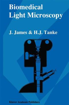
New interest in light microscopy of the last few years has not been backed up by adequate general literature. This book intends to fill the gap between specialized texts on detailed topics and general introductory booklets, mostly dealing with the use of the conventional light microscope only. In this short textbook both new developments in microscopy and basic facts of image formation will be treated, including often neglected topics such as axial resolving power, lens construction, photomicrography and correct use of phase-en interference contrast systems. Theoretical background will be dealt with as far as necessary for a well-considered application of these techniques enabling a deliberate choice for the approach of a certain problem. Over 150 illustrations (photomicrographs and diagrams) complete the information on microscopy of the nineties in the biomedical field, intended for scientists, doctors, technicians and research students. Many drawings have been contributed by the illustrator R. Kreuger; the photographic work has been executed by J. Peeterse. Secretarial assistance in preparing the manuscript was given by Ms T. M. S. Pierik. Dr M. J. Pearson has corrected the English of the final text.
1. Light microscopy as an optical system, the stand and its parts.- 1.1 Basic theory.- 1.2 The objective as an optical tool; resolving power.- 1.3 Eyepieces.- 1.4 Objective and eyepiece as an integrated system.- 1.4.1 The interplay between objective and eyepiece.- 1.4.2 Tube length.- 1.4.3 Axial resolving power and depth of field.- Recommended further reading.- 2. The light microscope as a tool for observation and measurement: illumination and image formation.- 2.1 Modulation of the illuminating light by the object.- 2.2 The stand and its parts.- 2.3 Illumination and image formation.- 2.3.1 General aspects.- 2.3.2 Types of illumination.- 2.3.3 Special types of illumination: darkground illumination.- 2.3.4 The light source.- 2.3.5 Confocal illumination.- Recommended further reading.- 3. Fluorescence microscopy.- 3.1 Theoretical background.- 3.1.1 What is fluorescence?.- 3.1.2 Physical properties of fluorescence.- 3.1.3 Spectral properties of fluorochromes.- 3.1.4 Quantum efficiency of fluorochromes.- 3.2 The fluorescence microscope.- 3.2.1 Incident or transmitted light illumination.- 3.2.2 Fluorescence microscopy with transmitted illumination.- 3.2.3 Fluorescence microscopy with incident illumination.- 3.2.4 Components of the fluorescence microscope.- 3.2.5 The two-wavelengths excitation method for fluorescence microscopy with incident light.- Recommended further reading.- 4. Special optical techniques of image formation.- 4.1 Phase-contrast microscopy.- 4.1.1 Basal theoretical facts.- 4.1.2 Practical realization of the phase-contrast.- 4.1.3 The phase-contrast image with different objects.- 4.2 Interferometry and interference contrast.- 4.2.1 Principles of image formation in interference contrast.- 4.2.2 Differential interference contrast.- 4.3 Modulation-contrast microscopy.- 4.4 Polarization microscopy.- 4.4.1 Anisotropy as an optical phenomenon.- 4.4.2 The polarized light microscope.- 4.5 Reflection microscopy and reflection-contrast microscopy.- 4.6 Acoustic microscopy.- 4.7 Superresolution: modern developments.- Recommended further reading.- 5. Reproduction of microscopic images, microphotography.- 5.1 Drawing and drawing apparatuses.- 5.2 Microprojection.- 5.3 Television microscopy.- 5.4 Photomicrography.- 5.4.1 Some basic principles.- 5.4.2 Photographic materials.- 5.4.3 Photomicrography in practice.- 5.4.4 Colour photomicrography.- 5.4.5 Photomicrography of fluorescence images.- 5.4.6 Special techniques in microphotography.- 5.4.7 Holographic photomicroscopy.- 5.4.8 Cinemicrography.- Recommended further reading.- 6. Quantitative analysis of microscopic images.- 6.1 Introduction.- 6.2 Morphometric techniques.- 6.2.1 Estimation of distances perpendicular to the optical axis.- 6.2.2 Measurements of distances along the optical axis.- 6.2.3 Measurements of surfaces and volumes: stereology.- 6.3 Counting methods.- 6.4 Absorption and fluorescence measurement of cells.- 6.5 Absorption cytophotometry (cytophotometry or microphotometry).- 6.5.1 Object plane scanners.- 6.5.2 Image plane scanners.- 6.6 Fluorescence cytophotometry (cytofluorometry, microfluorometry).- 6.6.1 Theoretical background.- 6.6.2 Practical aspects of cytofluorometry.- 6.7 Flow cytometry.- 6.8 Microspectrophotometry.- Recommended further reading.- 7. Automation: image analysis and pattern recognition.- 7.1 General introduction.- 7.2 Scanning of microscopic objects: special cameras.- 7.3 The digitized image.- 7.3.1 Image processing and image analysis.- 7.3.2 Spatial resolution and grey value resolution.- 7.3.3 Intensity transformations.- 7.3.4 Segmentation of images.- 7.4 Image analysis.- 7.5 Pattern recognition.- Recommended further reading.- 8. Appendix: technical aspects of the microscopical observation in practice.- 8.1 Introduction.- 8.2 Setting up a microscope for Köhler illumination.- 8.3 Again: the object.- 8.4 On the way through the object.- 8.5 Maintenance and minor technical problems.- 8.6 Frequently occurring minor defects.- Recommended further reading.- Index of subjects.

Date de parution :

Ouvrage de 192 p.

15.5x23.5 cm

Disponible chez l'éditeur (délai d'approvisionnement : 15 jours).

Prix indicatif 158,24 €

Ajouter au panier

Ces ouvrages sont susceptibles de vous intéresser


Video Microscopy
105,49 €