
Atlas of Scrotal Ultrasound, Softcover reprint of the original 1st ed. 1992
Langue : Français
Auteur : Martin Brigitte
Préfacier : Hricak H.
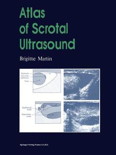
This unique ultrasound atlas is dedicated entirely to pathology of the scrotal contents. The 357 illustrations constitute the major proportion of the book, each of them is accompanied by a labelled diagram and detailed description. The atlas covers the essential embryology and ultrasound anatomy, the principal clinical presentations encountered in practice by the clinician and radiologists, and the differential diagnosis of intrascrotal cysts and calcifications. For each chapter there is a short but precise text, supplemented by tables or decision-making algorithms followed by the relevant images.
Embryology; technical notes and ultrasound anatomy.- The acute scrotum.- The chronically enlarged scrotum.- Impalpable testicular tumours within a clinically normal scrotal sac.- Male infertility.- Post-surgery scrotal sac.- Calcifications of structures within the scrotum and the periscrotal region.- Cystic structures of the scrotal contents.- Alphabetical index.
Date de parution : 08-2014
Ouvrage de 200 p.
21x27.9 cm
Thème d’Atlas of Scrotal Ultrasound :
Mots-clés :
diagnosis; fertility; hoden; infertility; scrotum; surgery; testis; ultraschall; ultrasound; Ultrasonography
© 2024 LAVOISIER S.A.S.