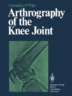
Arthrography of the Knee Joint, Softcover reprint of the original 1st ed. 1979
Langue : Anglais
Auteur : Thijn C.J.P.
Préfacier : Blickman J.R.

It is a great pleasure to introduce this book and its writer to the reader. Dr. Thijn has been interested in double contrast studies since he wrote his thesis on the double contrast examina tion of the colon. It would sound facetious to state that after he exhausted this field, he was in need of some other area where the same technique could be used. However, in the same exact and thorough way as in his colon studies, he has examined the knee joint. Considering that the knee is one of the most heavily taxed joints in man, with a multitude of afflictions, many of them closely connected with the age of the individual, radiological investiga tion has shown very few innovations over the decades. The true anteroposterior and lateral projections were ~ and still are ~ the mainstay of the investigation. Projections of the intercondylar fossa, and true patellar projections were used incidentally. Just prior to World War II the advent of arthrography as a double contrast investigation, as promoted by Oberholzer, was a real breakthrough.
1 History of Arthrography.- 2 Technique of Double Contrast Arthrography.- 2.1 Introduction.- 2.2 Injection of Contrast Media.- 2.3 Arthrography.- 2.3.1 Cruciate Ligaments.- 2.3.2 Menisci.- 2.3.3 Patellofemoral Joint.- 2.3.3.1 Lateral Projections.- 2.3.3.2 Tangential Projections.- 2.4 Aftercare and Complications of Arthrography.- 2.4.1 Hydrops.- 2.4.2 Allergic Reactions.- 2.4.3 Air Embolism.- 2.4.4 Arthritis.- 3 Meniscal Lesions.- 3.1 Specific Anatomy.- 3.1.1 Medial Meniscus.- 3.1.2 Lateral Meniscus.- 3.2 Meniscal Functions.- 3.3 Friction and Lubrication.- 3.3.1 Interface Lubrication.- 3.3.2 Buffer Lubrication.- 3.3.3 Elastohydrodynamic Lubrication.- 3.3.4 Sponge Lubrication.- 3.3.5 Lubrication by a Transient Increase in Viscosity.- 3.4 Etiology of Meniscal Lesions.- 3.4.1 Mobility of the Menisci.- 3.4.2 Risk-Increasing Factors.- 3.5 Normal Radiologic Anatomy of the Menisci.- 3.5.1 Medial Meniscus.- 3.5.2 Normal Lateral Meniscus.- 3.5.3 Spurious Meniscal Lesions.- 3.6 Meniscal Lesions.- 3.6.1 Meniscal Ruptures.- 3.6.1.1 Tangential Incisure.- 3.6.1.2 Longitudinal Ruptures.- 3.6.1.3 Fish Mouth Ruptures.- 3.6.1.4 Transverse Ruptures.- 3.6.1.5 Combined Ruptures.- 3.6.2 Types of Discoid Meniscus.- 3.6.3 Degeneration of Meniscal Cartilage.- 3.6.3.1 Primary Degeneration.- 3.6.3.2 Secondary Degeneration.- 3.6.4 State After Meniscectomy.- 3.7 Correlation Between Arthrography and Arthroscopy.- 4 Lesions of the Patellofemoral Joint.- 4.1 Anatomy and Physiology of the Patellofemoral Joint.- 4.2 Articular Cartilage.- 4.2.1 Histology.- 4.2.2 Nutrition of Cartilage.- 4.2.3 Properties of Cartilage.- 4.2.4 Cartilage Degeneration.- 4.2.5 Normal Radiologic Anatomy.- 4.3. Etiology of Patellar Chondropathy.- 4.3.1 General Aspects.- 4.3.2 Mechanical Lesions of the Patellar Cartilage.- 4.3.2.1 Exogenous Factors.- Direct Lesion of the Patella.- Overstress.- Fractures.- 4.3.2.2 Endogenous Factors.- Patellar Dysplasia.- Patella Partita.- Dysplasia of the Facies Patellaris Femoris.- High Patella.- Low Patella.- Increased Patellar Mobility.- Increased Pressure in the Patellofemoral Joint.- 4.3.3 Non-Mechanical Lesions of the Patellar Cartilage.- 4.4 Radiologic Diagnosis of Patellar Chondropathy.- 4.4.1 Radiologic Examination Without Contrast Medium.- 4.4.2 Double Contrast Arthrography.- 4.5 Correlation Between Double Contrast Arthrography and Arthroscopy.- 5 Cruciate Ligaments.- 5.1 Anatomy of the Cruciate Ligaments.- 5.2 Radiologic Technique.- 5.3 Radiologic Anatomy of the Cruciate Ligaments.- 5.3.1 Anterior Cruciate Ligament.- 5.3.2 Posterior Cruciate Ligament.- 5.4 Etiology of Cruciate Ligament Ruptures.- 5.5 Cruciate Ligament Pathology.- 5.5.1 Abnormal Delimitation of the Cruciate Ligaments.- 5.5.2 Abnormal Displacement of the Tibia in Relation to the Femur.- 5.5.3 Irregular Structures Within the Cruciate Ligament Compartments.- 5.6 Accuracy of Arthrographic Cruciate Ligament Diagnosis.- 6 Joint Capsule, Collateral Ligaments, Hoffa Body and Bursae.- 6.1 Capsule, Hoffa Body, and Collateral Ligaments.- 6.2 Bursae.- 6.2.1 Suprapatellar Bursa.- 6.2.2 Semimembranosogastrocnemial Bursa.- 6.2.3 Popliteal Bursa.- 7 Lesions of the Articular Cartilage.- 7.1 General Aspects.- 7.2 Etiology and Double Contrast Arthrography.- 7.2.1 Primary Form.- 7.2.2 Secondary Form.- 7.2.2.1 Direct Injury.- 7.2.2.2 Indirect Injury.- Meniscal Lesions.- Cruciate and Collateral Ligament Lesions.- Osteochondritis Dissecans.- Inflammations.- Changed Pressures.- Hemophilia.- Metabolic Disorders.- References.
Date de parution : 03-1979
Date de parution : 03-2012
Ouvrage de 158 p.
21x27.9 cm
Disponible chez l'éditeur (délai d'approvisionnement : 15 jours).
Prix indicatif 105,49 €
Ajouter au panierThème d’Arthrography of the Knee Joint :
© 2024 LAVOISIER S.A.S.


