Multimodal Retinal Imaging
Auteurs : Chopdar Amresh, Aung Tin
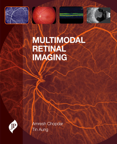
Retinal imaging techniques are used to diagnose and screen for various conditions such as macular degeneration and glaucoma. Equally importantly, they are used to monitor the progression of previously diagnosed conditions. In either case, the ability to interpret the results provided by the various techniques available to the clinician is of vital importance for the appropriate management of the underlying pathology.
Multimodal Retinal Imaging provides a timely summary of the various state of the art retinal imaging techniques now available and enables the reader to interpret the findings they will encounter in the care of patients with retinal conditions.
Section One TECHNIQUES, covers all the main modalities, such as Optical coherence tomography and Scanning Laser ophthalmoscopy and for each one:
- summarises the history and development of the technique in question
- explains relevant basic science (anatomy, physiology, etc)
- gives practical advice on using the technique in everyday practice (positioning of the patient, etc)
- provides a visual summary of the appearance of normal results
- highlights the appearance of the discrete abnormal signs that users of the technique should be aware of and alert to
Section Two DISORDERS covers the important pathologies relevant to retinal imaging, such as Retinal vascular disorders, and for each disease group:
- provides a brief overview of the disorders in question, their shared features and differentiating factors
- describes and illustrates the presentation of each specific condition via different modalities, showing multiple variations of each specific condition so that the reader can become familiar with the range of appearances likely to be seen in practice.
Key Points
- Provides unique Technique and Disease-based approach to provide readers with comprehensive but highly practical clinical reference to retinal imaging
- Includes information on key areas of recent research and development such as imaging of the outer retina and choroid and retinal vasculature imaging (including vessel calibre, branching and fractals)
1 Fundamentals of fluorescence angiography
2 Indocyanine angiography
3 Optical coherence tomography (OCT) and Scanning Laser Ophthalmoscopy (SLO)
4 Ocular echography
5 Autofluorescence
6 Optic nerve head imaging
7 Fundamentals of multimodality imaging
8 Retinal vascular disorders
9 Macular degeneration
10 Macular dystrophies
11 Choroidal disorders
12 Optic nerve disorders
13 Intraocular neoplasms
14 Diabetic retinopathy
15 Glaucoma
Amresh Chopdar, FRCOphth
Consultant Ophthalmologist, Surrey & Sussex NHS Trust, East Surrey Hospital, Redhill, UK
Tin Aung, FRCOphth, FAMS,PhD(Lond)
Deputy Director, Singapore Eye Research Institute, Associate Professor, Dept of Ophthalmology, Yong Loo Lin School of Medicine, National University of Singapore
Date de parution : 08-2014
Ouvrage de 173 p.
21.9x27.5 cm
Disponible chez l'éditeur (délai d'approvisionnement : 14 jours).
Prix indicatif 154,17 €
Ajouter au panier